ISSN : 0975-5276
EISSN : 0975-9174

SURESHA H.R.1, KRISHNAPPA M.2*, DESCALS E.3, RAJU G.H.4, TAYLOR B.R.5
1Department of P.G. Studies and Research in Applied Botany, Kuvempu University, Shankaraghatta- 577 451, Shivamogga, Karnataka, India.
2Department of P.G. Studies and Research in Applied Botany, Kuvempu University, Shankaraghatta- 577 451, Shivamogga, Karnataka, India.
3Instituto Mediterráneo de Estudios Avanzados, Calle Maestro Miquel Marquès, 21,07190 Esporles, Mallorca, Balears, Spain.
4Department of Biology, St. Francis Xavier University, 2320 Notre Dame Avenue, P.O. Box 5000, Antigonish, Nova Scotia, Canada B2G 2W5.
5Department of Biology, St. Francis Xavier University, 2320 Notre Dame Avenue, P.O. Box 5000, Antigonish, Nova Scotia, Canada B2G 2W5.
* Corresponding Author : krishnappam4281@yahoo.com
Received : 08-09-2012 Accepted : 03-04-2013 Published : 08-04-2013
Volume : 5 Issue : 3 Pages : 410 - 416
Int J Microbiol Res 5.3 (2013):410-416
DOI : http://dx.doi.org/10.9735/0975-5276.5.3.410-416
Aquatic ecosystems include both fresh and marine water bodies comprising both abiotic and biotic components. The biotic component consists of producers, consumers and decomposers. Microorganisms play an important role in decomposition of organic matter; in fresh water much of this decomposition is carried out by a group of Deuteromycete fungi known as aquatic hyphomycetes. The diversity of aquatic hyphomycetes in a river can be assessed indirectly because they produce distinctively shaped conidia that persist in the water and are often concentrated in foam. Production of conidia may also be induced by incubating decomposing organic matter. The present study was carried out to explore the diversity of aquatic hyphomycetes on leaf bits in Kalathgiri Falls, in Chikmagalur District of Karnataka. The fallen leaves of four species of riverside plants were tied in nylon mesh bags, incubated in the stream for 15 days, and examined in the laboratory for spores indicating the presence of aquatic fungi. A total of 18 species of aquatic hyphomycetes belonging to 13 genera were recorded. There was little evidence of species associations, and the assemblage at each site along the falls was distinct.
Hyphomycetes, Conidia, Diversity, Aquatic fungi, Decomposition.
A knowledge of general diversity of the native mycota is most important when considering that each fungal species has its own niche in the habitat. Fungi play a major role in the food web as organic matter decomposers and contributors to nutrients cycling because they degrade aquatic dead animals and plants. Most filamentous fungi in streams are members of the aquatic hyphomycetes or Ingoldian fungi, a group of Ascomycota and Basidiomycota which reproduce asexually by spores (conidia) [1-4] . The main habitats of these aquatic fungi are streams, reservoirs, lakes and waterfalls, on substrates like leaves, twigs and branches of trees. However they also occur to some extent on submerged, decaying culms of the softer reeds in lakes.
Hyphomycetes are an important group in the aquatic community because they degrade leaves and twigs in water [5-7] . Leaf breakdown in streams is caused by the action of physical factors, the activity of detritus-feeding macroinvertebrates called shredders [8] and the activity of microorganisms such as aquatic hyphomycetes [1,7] . Aquatic hyphomycetes are the main microbial decomposers of leaf litter in streams [7,9] . They occupy the base of the decomposer food chain, and therefore influence all other stream organisms by sequestering or releasing nutrients, providing a direct food source for leaf-shredding insects, releasing fine particulate organic matter that is consumed by other invertebrates, and providing energy to support the entire grazing food chain up to fish [8] . Aquatic hypomycetes have been studied mainly on leaves and needles of riparian trees [7,8] , or sometimes fallen wood [10] .
Diversity of hyphomycetes has been correlated with temperature [11] , water chemistry [12] and quality of riparian vegetation [13] Temperature affects their growth and sporulation [11,14,15] and light acts as a stimulus for their sporulation on leaf litter [16-18] Fabre [13] in his detailed study suggests that altitude is an important factor in structuring aquatic hypomycete communities in streams of southwestern France. Suberkropp and Klugg [7] and Singh and Mosa [19] distinguished warm season and cold season fungi. Changes in chemical composition during decomposition of submerged plant materials show selective inhibition or stimulation of fungal species and biological interactions, resulting in a succession of fungal species [10] .
Aquatic fungi produce conidia abundantly in the aquatic environment and even in terrestrial water among soil particles [19] . Hyphomycete fungi can rapidly colonize available substrates within a few days, thereafter releasing prodigious numbers of conidia [6,9] , which disperse throughout the water column. Hence, filtration of stream water is an effective method for trapping and concentrating hyphomycete conidia [3,20] . Conidia tend to concentrate on the surface of air bubbles, which under certain conditions, especially if the water is rich in dissolved organic matter, congregate as persistent foam [21] . Individual species of hyphomycetes can be identified by the distinctive and often complex shapes of their conidia, although convergent evolution has led to many similar forms [1] .
Several studies on the diversity of freshwater fungi, based on captured spores, have previously been carried out in temperate and subtropical regions [1,20,22,23] and a significant number of new species have been discovered in the last ten years alone [17] . Many species appear to have a world-wide distribution, while others may be restricted to warmer or cooler climates. Further sampling is needed to better establish the distributions of these fungi, especially in less well studied tropical and sub-tropical regions. The stream bed of low order, tropical streams is composed mainly of dead leaves and tree branches originating from the surrounding forest. Leaf debris is therefore the logical place to search for aquatic hyphomycetes. In the present study colonization of aquatic hyphomycete fungi at Kalathgiri Falls, Chikmagalur District, was studied by examining spores produced on leaf debris.
Kalathgiri Falls, located at 13° 32′ 56" N latitude and 75° 47′ 20" E longitude, comes under Chikmagalur District of the western part of Karnataka State in India [Fig-1] . The total area of Chickmagalur comprises 7201 km2 of which 58% is forested. The temperature varies from 15°C to 28°C and the annual rain fall is 1900 mm. The heavy rain fall has resulted in many water tanks, streams and reservoirs. The Kalathgiri waterfalls have flowing water throughout the year and are surrounded by thick vegetation. Abundant foam develops in the water in all seasons. Four study sites, labelled A, B, C and D, were established at Kalathgeri Falls, each of which was further sub-divided into two (A1A2, B1B2, C1C2, D1D2).
Leaf litter from four species of vegetation common around Kalathgiri Falls was used as substrate to attract aquatic hyphomycetes. The litter types were bamboo, Pongamia glabra, Tectona grandis and Terminalia sp. Leaves from each species were collected and cut into small pieces. Mesh bags containing leaf fragments were submerged in moving water at each site. Bags were removed at 5-day intervals for 30 days and aquatic hyphomycetes were isolated from leaf fragments in each bag.
During the investigation, foam samples were collected from lotic water bodies of the study area. Foam samples were also collected in forests. Foam samples were collected in small plastic bottles and sterile polythene bags, which were also used to collect leaf debris. The substrate of plant leaves and wood pieces were also kept for incubation and fungi were isolated by a plating technique. When monocultures were growing, they were further studied for their colony characteristics. These fungi were further mass multiplied by the help of a baiting technique and were stored in sterile polyethythene bags.
The hair technique was used to capture single spores in isolation under the dissecting microscope. The incubated plates were placed under the microscope. The conidial expression was observed, and after confirming that conidia were being released, single conidia were transferred on a human hair to malt extract plates. The plates were incubated at 18°C for one week. The micropipette technique was used to capture floating spores in incubated plates. The incubated plates were placed under the dissecting microscope and the conidia were observed. The selected spores were sucked into a micropipette and directly transferred to the malt extract plate.
A cross-section of the spore types observed at Kalathgiri Falls is illustrated in [Plates-1a] , [Plates-1b] , [Plates-1c] , [Plates-1d] , [Plates-1e] and [Plates-2a] , [Plates-2b] , [Plates-2c] , [Plates-2d] , [Plates-2e] . These species are described below.
Habitat: Foam samples and decaying leaf samples
Conidia: Conidia are sigmoid and very long and thin, up to 300 x 5-7 µm. A. furtiva, which is 50% longer than A. longissima, was recognized as a separate species only recently (1999).
Remark: This species is rare at Kalathgiri Falls, but is common and widespread in temperate zones.
Habitat: Conidia in foam samples
Conidia: The distinguishing feature of these sinusoidal conidia is their enormous size, 500-700 µm length [17] .
Remark: This species appears to be largely tropical in distribution. It has been reported in Pakistan [23] and Hawaii [25] but is not included among common species in Eurasia [2] and North America [1] .
Habitat: Conidia found in foam samples
Conidia: Conidia are tetraradiate, narrowing to a long tip, the basal arm slightly larger than the three terminal arms, which are markedly restricted at the point of attachment.
Remark: The conidia in this type of species is common in many parts of the world, especially in temperate regions. Its presence in India has been confirmed in pure cultures derived from conidia in foam samples.
Habitat: Conidia in leaf sample
Conidia: The conidia of this widespread species are very large (>500 µm) and complex. They consist of a long central axis from the base of which arise two sets of long, thin (4-5 µm) axial branches, often in whorls. The axial branches may themselves sometimes be branched, making the overall shape of the spore rather variable. Although well known and common in temperate zones [1-3] , it appears to be less common in the tropics.
Remark: This organism is reported for the first time in Chickmagalur District.
Habitat: Conidia in leaf sample
Conidia: This distinctive species has seven long, thin arms, equal in length, radiating from the short stalk of the conidium, giving the spore a stellate or multiradiate appearance. F. verticillata is evidently a tropical species; it was first described from Nigeria [26] , but it is absent from Europe (British Mycological Society Checklist of Fungal Names, 2012) and North America [1] .
Remark: This organism is reported for the first time in Chickmaglur District.
Habitat: Conidia in foam samples
Conidia: The conidia have three arms, equal in length (50-100 µm), arising from a basal stock which makes a fourth arm, sometimes shorter. This species is commonly found in the rainy season.
Remark: The conidia of this temperate-zone species, uncommon at our sites, have also been found in some regions of the Western Ghats, and in the Sinharaja Forest of Sri Lanka [22] .
Habitat: This organism is found in leaf samples
Conidia: As the Latin name suggests, the elongate spores of this species are curved like a crescent moon. There is a persistant attachment scar on the outside curve, proximal to the end of the conidium.
Remark: Ingold [3] notes that this species is worldwide in distribution but appears to be more abundant in the tropics. It was found at many sites in Sri Lanka [22] .
Habitat: Conidia in foam sample
Conidia: The spores are long, ~100 µm, branched and gracefully curved. The central axis bends in the middle, from which region arise two more branches of equal length, creating a tetraradiate conidium. All the connections are unrestricted and the branches are all equally thin.
Remark: This organism is reported for the first time in Chikmagalur District.
Habitat: Conidia found in leaf sample
Conidia: The conidia are unusually simple among aquatic hyphomycetes: they are elongately ellipsoidal, tapering narrowly at both ends, and the divisions between three internal cells are conspicuous.
Remark: This species was originally known as Dactylella aquatica [3] . Although common in temperate climates [1,3] it was rare at Kalathgiri Falls.
Habitat: Conidia found in leaf sample.
Conidia: It is tetra-radiate, with each of the branches narrowing where it meets the main axis. The main axis also extends past the union of the three branches, making the conidium easy to distinguish.
Remark: Ingold [16] notes that this species is abundant in tropical countries, and also widespread, occurring essentially worldwide.
The decaying leaves of all four tree species collected from Kalathgiri Falls revealed a total of 18 species of aquatic hyphomycetes belonging to 13 genera, among which Anguillospora crassa was dominant overall. Fungal sporulation was most abundant along the surface of bamboo leaves, which supported eight species of hyphomycetes. On the leaves of Pongamia glabra, six species of aquatic hyphomycetes were observed, among which, Flagellospora curvula was dominant. The leaves of Tectona grandis revealed six hyphomycete species, among which Triscelophorus monosporus was dominant. Leaves of Terminalia sp. also expressed six species of aquatic hyphomycetes, dominated by Lemonniera aquatica. Therefore, fungal communities on each litter type differed both in the number of species they contained and in the dominant species. Overall, leaves collected in the margin of Kalathgiri Falls showed dominance of Anguillospora spp.
Previous studies indicate that when a specific substratum is introduced into the falls, fungal species richness is initially low, rises to a peak within a few weeks, then remains stable or declines slightly. Two plant materials baited in these experiments (Pongamia glabra and Terminelia sp.) also show this basic pattern, maintaining or slightly increasing their species richness of hyphomyctes for the first 10 days of immersion, before a long decline thereafter. On the other two substrates, bamboo and Tectona grandis, species richness declined steadily from the outset [Fig-2] .
Overall, species richness on all four litter types decreased substantially from 5 to 30 days in the water, probably following the advancing decay of the leaves.
These plant leaves revealed that aquatic fungal diversity changes according to the particular type of litter being colonized. The list of aquatic hyphomycetes given in [Table-1] records their occurrence in foam samples and on decaying plant material collected at different localities whenever the seasonal species arose. Species whose conidia were observed only sporadically and whose identities were Anguillospora crassa was the most widespread species, occurring at seven of eight sites. Triscelophorus monosporus occurred at six sites, and Alatospora accuminata occurred at five sites. All the remaining species were occasional to rare, occurring at 1‑4 sites in no clear pattern [Table-1] . Species richness at individual sites ranged from 11 at Site A1 to only four at Site D2. At the level of the whole falls, the hyphomycete community appears to be composed of a few widespread species and a large number of occasional or transitory species.
There was little uniformity in species composition within main sites: Sites A1 and A2 shared three species in common, while the remaining site pairs shared only one or two species [Table-1] . Hence community composition showed very little similarity within main sites; Jaccard Coefficient ranged from 0.23 (Site A1 vs. Site A2) to 0.06 (Site B1 vs. B2). Comparing all sites, the hyphomycete assemblages at sub-sites C1 and D1 were quite similar (Jaccard Coefficient 0.78) but all other site combinations were different.
Similarly, there were few strong species associations among sites. Articulospora tetracladia and Dendrospora erecta always occurred together (four sub-sites), as did Flagellospora curvula with Tetrachaetum elegans (three sub-sites) and Lunulospora curvula with Tumularia aquatica (B2 only). Cluster analysis recognized one group of fungi that comprised three common species (An. crassa, Al.accuminata, L. aquatica) plus Clavariopsis aquatica (Jaccard Coefficient 0.71) and a second that combined uncommon Flagellospora curvula and T. elegans with Lemonniera filiformis (0.75). None of the other species defined strong clusters. At the local level, it appears that each location has a more or less unique assemblage of fungi which has little overlap with places nearby. This result suggests a degree of site selection on the part of the fungi, perhaps based on the substrates (leaves and wood) available at each location.
Total abundance of each species was recorded at the eight study sites at Kalathgiri Falls. The eighteen species are arranged in order of relative abundance in [Fig-3] . Shannon-Weaver index for this assemblage is 2.80, indicating a relatively diverse community. Although Anguillospora crassa was both the most frequent species [Table-1] and the most abundant [Fig-3] , other species that occurred at only a few sites were relatively abundant where they were found. These results indicate that some species of aquatic hyphomycetes can tolerate adverse environmental conditions and thrive in habitats that are stressful to other species. There is clearly a strong selection for particular substrata such as plant leaves, where different fungal species may be dominant on different litter types.
The trend of decreasing abundance with increasing species rank in [Fig-3] fits well to a negative exponential model (R2 = 0.91) with a relatively shallow slope; that is, abundance declines rather slowly from the most common to the least common species in the community. By extrapolating this line to abundance of zero, an estimate of the total number of species in the community of 32.2 is obtained. Thus, we may expect up to 14 additional, rare species may be present at Kalathgiri Falls. This pattern of one or a few dominant species (in this system, Anguillospora crassa) a larger group of frequent species, and a “long tail†of rare species has been frequently observed in aquatic fungi [27,28] .
Seasonal variation and other factors (biotic and abiotic) connected with a stream, particularly alkalinity, ion concentrations, pH and temperature that vary seasonally and that may affect fungal species richness and distribution [1] have not been considered here. However, characteristic assemblages of these fungi have been studied worldwide by various workers. It is clear from these studies that the distribution of certain species is related to temperature in that major differences in species composition have been noted between temperate and tropical streams [4] . The occurrence of individual species in the Chikmaglur District was different from those recorded in the temperate regions. For examples, Tetrachaetum elegans and Tumularia aquatica are largely tropical and sub-tropical in distribution, while Anguillospora crassa, Lunulospora curvula and Triscelophorus monosporus, are more widespread and commonly occurring.
Gunasekera & Rukmani [22] surveyed aquatic hyphomycete spores in 15 streams throughout Sri Lanka, in the same climatic region as the current work. They identified 15 species, of which nine are identical to species observed at Kalathgiri Falls (Jaccard Coefficient = 0.38). Hence, while there appears to be great specificity in species assemblages at the site level, many species are apparently common throughout the broader geographic region. However, while the second-most frequent species in both Sri Lanka and Kalathgiri Falls was the same (Triscelophorus monosporus), the most frequent species in Sri Lanka was Lunulospora curvula (15 of 15 sites), which was observed only once here.
A diveristy of aquatic hyphomycetes rapidly colonized submerged leaf litter at Kalathgiri Falls. Peak species richness of 4-8 species occurs within the first week after litter is submerged, declining thereafter as the substrate decomposes. The species assemblage, as indexed by spore production, varies at the local level according to charactertistics of a particular site, the litter type being colonized, and the stage of decomposition of that litter. Hence, the composition of the spore assemblage sampled varies widely through space and time even within Kalathgiri Falls. Abundances of most fungal species appear to vary more or less independently of other species, leading to only limited co-occurrences and no evidence of recognizable species associations. Nevertheless, many of these species are widespread or cosmopolitan at the regional level and significant overlap with species lists from other sites in the same geographic and climatic zone are likely.
Sampling of the fungal community based on conidia is naturally biased by the differential spore production among species, and their unequal tendencies to persist in the water or become concentrated in foam. Conidia sampling will therefore always underestimate the true diversity of the fungal community. Species abundance ranking suggests that roughly half the species of aquatic hyphomycetes present at Kalathgiri Falls were observed, the remainder presumably being represented by rare species or those whose spores elude collection. A few unidentified spores probably belong to this group of missed species.
The first author is very thankful to the Department of Applied Botany, Kuvempu University, which provided the laboratory facilities for his work. The fourth author extends thanks to Dr. Felix Bärlocher for help with hyphomycete taxonomy.
[1] Bärlocher F. and Marvanová L. (2010) Assessment of Species Diversity in the Atlantic Maritime Ecozone, NRC Research Press, Ottawa, Canada. 1-37.
» CrossRef » Google Scholar » PubMed » DOAJ » CAS » Scopus
[2] Gulis V., Marvanová L. and Descals E. (2005) Methods to Study Litter Decomposition: A Practical Guide, Springer, The Netherlands, 153-167.
» CrossRef » Google Scholar » PubMed » DOAJ » CAS » Scopus
[3] Ingold C.T. (1975) An Illustrated Guide to Aquatic and Water-Borne Hyphomyctes (Fungi Imperfecti) with Notes on Their Biology, Scientific Publication, Freshwater Biological Association, Ambleside, UK, 96.
» CrossRef » Google Scholar » PubMed » DOAJ » CAS » Scopus
[4] Nilsson S. (1964) Symbolae Botanicae Upsalienses, Institute of Systematic Botany, Uppsala, Sweden, XVIII, 2, 1-130.
» CrossRef » Google Scholar » PubMed » DOAJ » CAS » Scopus
[5] Kaushik N.K. and Hynes H.B.N. (1971) Archiv fur Hydrobiologia, 68, 465-515.
» CrossRef » Google Scholar » PubMed » DOAJ » CAS » Scopus
[6] Suberkropp K. (1991) Mycological Research, 95, 843-850.
» CrossRef » Google Scholar » PubMed » DOAJ » CAS » Scopus
[7] Suberkropp K. and Klug M.J. (1981) The Fungal Community, its Organization and Role in the Ecosystem, Marcel Dekker Inc., New York, USA, 761-776.
» CrossRef » Google Scholar » PubMed » DOAJ » CAS » Scopus
[8] Cummins K.W. (1974) BioScience, 24, 631-641.
» CrossRef » Google Scholar » PubMed » DOAJ » CAS » Scopus
[9] Bärlocher F. (1992) Freshwater Biology, 28, 1-7.
» CrossRef » Google Scholar » PubMed » DOAJ » CAS » Scopus
[10] Shearer C.A. and Webster J. (1991) Mycological Research, 95, 413-420.
» CrossRef » Google Scholar » PubMed » DOAJ » CAS » Scopus
[11] Chauvet E. and Suberkropp K. (1998) Applied and Environmental Microbiology, 54, 1522-1525.
» CrossRef » Google Scholar » PubMed » DOAJ » CAS » Scopus
[12] Suberkropp K. (1998) Mycological Research, 102, 998-1002.
» CrossRef » Google Scholar » PubMed » DOAJ » CAS » Scopus
[13] Fabre E. (1996) Comptes Rendus de l’Academie de Sciences, Paris. Sciences de la Vie, 319, 107-111.
» CrossRef » Google Scholar » PubMed » DOAJ » CAS » Scopus
[14] Koske R.E. and Duncan I.W. (1974) Canadian Journal of Botany, 52, 1387-1391.
» CrossRef » Google Scholar » PubMed » DOAJ » CAS » Scopus
[15] Suberkropp K. (1984) Transactions of the British Mycological Society, 82, 5.
» CrossRef » Google Scholar » PubMed » DOAJ » CAS » Scopus
[16] Fisher P.J. and Webster J. (1978) Transactions of the British Mycological Society, 71, 465-478.
» CrossRef » Google Scholar » PubMed » DOAJ » CAS » Scopus
[17] Rahashekhar M. and Kaveriappa K.M. (2000) Hydrobiologia, 441, 149-153.
» CrossRef » Google Scholar » PubMed » DOAJ » CAS » Scopus
[18] Thomas K., Chilvers G.A. and Norries R.H. (1991) Mycological Research, 95, 178-183.
» CrossRef » Google Scholar » PubMed » DOAJ » CAS » Scopus
[19] Singh N. and Mosa T.M (1977) Transactions of the British Mycological Society, 68, 103-106.
» CrossRef » Google Scholar » PubMed » DOAJ » CAS » Scopus
[20] Bärlocher F. (1987) Canadian Journal of Botany, 65, 76-79.
» CrossRef » Google Scholar » PubMed » DOAJ » CAS » Scopus
[21] Ingold C.T. (1975) Transactions of the British Mycological Society, 65, 522-527.
» CrossRef » Google Scholar » PubMed » DOAJ » CAS » Scopus
[22] Gunasekera S.A. and Rukmani M.P.R. (1984) Journal of the National Science Council of Sri Lanka, 12, 273-282.
» CrossRef » Google Scholar » PubMed » DOAJ » CAS » Scopus
[23] Iqbal S.H. (1994) Mycoscience, 35, 331-343.
» CrossRef » Google Scholar » PubMed » DOAJ » CAS » Scopus
[24] Shearer C.A. and Descals E. (2007) Biodiversity Conservation, 16, 49-67.
» CrossRef » Google Scholar » PubMed » DOAJ » CAS » Scopus
[25] Anastasiou C.J. (1964) Pacific Science, 18, 202-206.
» CrossRef » Google Scholar » PubMed » DOAJ » CAS » Scopus
[26] Alasoadura S.O. (1968) Nova Hedwigia, 15, 419-421.
» CrossRef » Google Scholar » PubMed » DOAJ » CAS » Scopus
[27] Fierer N. (2008) Assessing Uncultivated Micro-organisms: From the Environment to Organisms and Genomes and Back, ASM Press, Washington, D.C., 95-110.
» CrossRef » Google Scholar » PubMed » DOAJ » CAS » Scopus
[28] Fróhlich J., Hyde K.D. and Petrini O. (2000) Mycological Research, 10, 1202-1212.
» CrossRef » Google Scholar » PubMed » DOAJ » CAS » Scopus
 | Fig. 1- Map of Chikmagalur District, Karnataka, India |
 | Fig. 2- Comparison of sporulation of aquatic hyphomycetes among litter types in incubation studies |
 | Fig. 3- Relative abundances of aquatic hyphomycete spores among species at Kalathgiri Falls. Names of numbered species are given in [Table-1]. |
 | Plate 1a- Anguillospora gigantea |
 | Plate 1b- Anguillospora furtiva |
 | Plate 1c- Articulospora tetracladia |
 | Plate 1d- Dendrospora erecta |
 | Plate 1e- Flabellospora verticillata |
 | Plate 2a- Lemonniera aquatic |
 | Plate 2b- Lunulospora curvula |
 | Plate 2c- Tetrachaetum elegans |
 | Plate 2d- Tumularia aquatica |
 | Plate 2e- Triscelophorus monosporus |
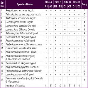 | Table 1- Occurrence of aquatic fungi in Kalthgiri Falls, based on spores collected from foam and decaying plant material |