


ISSN : 0976-8769
EISSN : 0976-8777



TALOUIZTE H.1, MERZOUKI M.2, BENLEMLIH M.3, AMRAOUI B.M.4*
1Laboratory of Vegetal and Urban Environmental Sciences, Ecole Normale Supérieure de Fès, PO Box 5206 Bensouda Fez,University Sidi Mohamed Ben Abdellah Fez, Morocco.
2Laboratory of Biotechnology, Department of Biology, Faculty of Sciences Dhar El Mehraz, PO Box 1796 Atlas, University Sidi Mohamed Ben Abdellah Fez, Morocco.
3Laboratory of Biotechnology, Department of Biology, Faculty of Sciences Dhar El Mehraz, PO Box 1796 Atlas, University Sidi Mohamed Ben Abdellah Fez, Morocco.
4Laboratory of Vegetal and Urban Environmental Sciences, Ecole Normale Supérieure de Fès, PO Box 5206 Bensouda Fez,University Sidi Mohamed Ben Abdellah Fez, Morocco.
* Corresponding Author : mbamraoui@yahoo.fr
Received : 20-09-2013 Accepted : 12-11-2013 Published : 15-11-2013
Volume : 3 Issue : 1 Pages : 35 - 41
J Toxicol Res 3.1 (2013):35-41
In this study, we evaluated the genotoxic effects of textile effluents on primary (PR) and secondary (SR) roots of <i>Vicia faba</i> L., <i>Allium sativum</i> L. and <i>Lactuca sativa</i> L. using cytogenetic tests. Genotoxicity was estimated in terms of micronuclei (MN) and mitotic index (MI) frequencies. Our results demonstrated that textile effluent reduced significantly seed germination, seedling growth and water status. Cytogenetic tests showed great differences between species in the response of both types of roots to the effluent. The differences are observed in MI frequency and suggested that, contrary to <i>Lactuca, Vicia</i> and <i>Allium</i> SR were more resistant to textile effluent than PR, whereas differences in MN indicated that, in contrast to <i>Vicia,</i> PR of <i>Allium</i> and <i>Lactuca</i> were more resistant to the effluent than SR. Thus, this study demonstrated that (i) the mechanism of mitotic activity is totally independent of that of micronucleus formation but are time consistent under effluent stress (ii) the response mechanisms of PR meristematic cell chromosomes to the effluent are different from those of SR. It has also been concluded that textile effluent affected PR cell chromosomes essentially by inhibition of mitosis while those of SR by nucleus fragmentation.
Textile effluent, plant species, roots, genotoxicity
Textile industry is one of the most polluting activities generating huge quantities of wastewater containing large groups of chemicals and residues of synthetic dyes which have caused serious environmental problems [1] . In Morocco, textile like several other industries generates a number of pollutants, which are discharged into the environment without any treatment. The effluents from textile industries are characterized by chlorides, residual chlorine and heavy metal pollutants [2] but contain also high level of azo dyes [3] , which are toxic, highly persistent because they have complex aromatic structures which make them highly resistant to degradation and thus may cause severe environmental problems. Large amounts of structurally different dyes are used for textile dyeing and around 10 to 15% of all dye is directly lost to wastewater [4] .
There are few studies in the literature on the evaluation of the hazards of textile effluents on higher plants. Earlier works have shown that action of organic contaminants on plant cells during exposure become initially noticeable with changes in the configuration of the nucleus. Simultaneously inhibition of DNA synthesis takes place with damage of the barrier function of the plasmalemma and its ability to accumulate calcium [5,6] . Other studies have examined the long term effect of these compounds but only on the cytoplasm, the vacuole and cell organelles. In contrast, there are few studies in the literature about the genotoxic effect of textile effluents or dyes on different plant organs. According to our knowledge, there are no studies on the chromosomal behavior of different parts of the root system which is more exposed to the toxicity of textile effluents. Less attention is given to the response of different types of roots to textile effluents, even less to the mechanisms involved in their resistance and in particular to the chromosomal mechanisms of primary and secondary plant roots.
With this background, we investigated the effects of textile effluent on seed germination and growth of Lactuca sativa L. On the other hand, the cytotoxicity, phytotoxicity and genotoxicity tests have been performed on Allium sativum L., Vicia faba L. and Lactuca sativa L. to assess the toxic effects of contaminants present in textile effluents on primary and secondary root meristematic cells.
The real textile effluent used in this study was obtained from a local textile industry of Sidi Brahim industrial district, Fez-Morocco. Various physico-chemical characteristics of textile effluent were analyzed using standard methods [7] . The textile dye Direct Green BL (C.I Direct Green 26, M.W. = 1333.1 g/mol) used in dye diffusion experiment was obtained as a gift from the same textile factory.
The organic compounds contained in real textile effluent samples were extracted with pure dichloromethane. GC/MS analysis of the extracts was performed on a Thermo Polaris Q mass spectrometer linked to a TRACE GC Ultra gas chromatograph equipped with Rtx®-5MS fused silica capillary column (30 m x 0.25 mm x 0.25 µm). Operating conditions were as follows: Temperature: Injector 250°C; Detector 200°C; Oven 250°C; Carrier gas: nitrogen with a flow rate of 1 ml/min. The volume injected was 1 µl. The peaks were identified by comparing their retention times with those of standards.
Seeds of lettuce (L. sativa L.) were placed in sterilized glass Petri dishes with two filter paper discs moistened with 5 ml of distilled water (control) or textile effluent and incubated at 25°C. The number of germinated seeds was counted, and expressed as the percentage germination, with respect to control. All the experiments were carried out in triplicate and the results were averaged.
Pots of 8 cm (diameter) × 12 cm (height) size were filled with equal amounts of see sand and ten 3 days old seedlings of L. sativa L. were planted in each pot. The plants were irrigated with distilled water (control) or with textile effluent once a day and grown under controlled conditions (16:8, light:dark photoperiod; day-time temperature: 25°C and 18°C at night). At the end of experiment, 15 day plants were collected. Root length, fresh weight and dry weight (after 2 days in an oven at 70°C) were recorded.
Seeds of lettuce (L. sativa L.) were germinated in plastic tray with two filter papers moistened with distilled water and incubated at 25°C. After 3 days of germination, the seedlings were placed in watch glasses containing solutions of Direct Green BL dye with a concentration of 100 mg/l, at pH of 3.5, 4.5 and 9.5, and incubated at 20°C during one hour [Table-1] . After incubation, root cells status was visualized using a DM-15 Optika microscope.
The genotoxic effect of real textile wastewater was examined by using the micronucleus test. Root tip micronucleus assay was carried out on three different plant species: Vicia faba L., Allium sativum L. and Lactuca sativa L. Seeds of tested species were germinated in filter paper at 25°C (V. faba L. and L. sativa L.) or small tubes (A. sativum L. gloves) containing tap water (control) or real textile effluent during three, seven and nine days [Fig-1] .
Cytological analysis were performed on the same treated seedlings from which primary root tips were taken after three days treatment and then secondary root tips after seven and nine days of exposition to effluent. Root tips of the three tested plant species were fixed in Carnoy’s fixative (absolute ethanol/acetic acid 3:1) for 24 h. Then fixative solution was removed and the material was stained according to Feulgen’s staining protocol [8] . Five root tips per experimental group were used for preparing slides. For micronucleus assay 1000 nuclei per slide were analyzed using photonic microscope: the MN frequency was compared with that of control, moreover MI (number of mitosis per 100 nuclei) was determined.
The results of genotoxicity assays on root tip apices of Vicia, Allium and Lactuca, were obtained from the examination of five different samples (1000 nuclei per slide) and statistically analyzed by Student’s t-test, p ≤ 0.05. The results were expressed as means ± s.d. (standard deviation).
The physico-chemical characterization of real textile effluent used in this study is shown in [Table-2] . The textile wastewater is characterized by high salinity and high SS content reflected by a TDS concentration of 3131 mg/l, an EC of 24.5 mS/cm and a SS concentration of 2810 mg/l. The concentration of COD and color measured by the absorbance of the effluent at λmax= 606 nm were 887 mg/l and 0.658 respectively. The color of the effluent was dark blue. N-NO3-, N-NO2-, N-NH4+ and P-PO43- concentrations were 10.46, 0.95, 0.96 and 1.15 mg/l, respectively. Very low biodegradability was observed, with BOD5/COD value of 0.13.
Finally, pH was quite neutral and averaged 7.27. The organic pollutants present in textile effluent were determined with GC/MS. Large amounts of organic compounds such as esters, alkanes and benzenes were identified using NIST library and listed in [Table-3] .
Textile effluent reduced significantly germination, root length and growth of Lactuca sativa L. seedlings [Table-4] . The germination percentage of lettuce seeds in textile effluent (64%) was lower than in tap water (control, 100%) and persisted up 3 days.
The Direct Green BL dye is detected after 1 h in the apoplasm, the cell membrane and the nucleus [Fig-2] (a), (b) & (c). We have noticed that Direct Green BL dye at pH 3.5 diffuse through root hairs [Fig-2] (b) but accumulate more in the nucleus and produce an intense invagination of the cytoplasmic membrane at pH 4.5 [Fig-2] (d). In contrast, the dye crossed low the membrane barrier at pH 9.5 [Fig-2] (e). Moreover, when textile effluent was applied 3 days to lettuce since the beginning of seed germination, the cytoplasm and vacuole in the cell layers of the PR tip area become heavily stained with the dyes present in textile effluent [Fig-2] (f).
The genotoxic effects of textile effluent were evaluated by means of cytogenetic analyses considering MN assay and MI values on root tips of three different plant species: V. faba L., A. sativum L. and L. sativa L., after 3 days (primary roots only), 7 and 9 days (secondary roots) of exposure. After 3 days of textile effluent exposure, mitotic activity of V. faba L. and A. sativum L. PR was heavily reduced from 19.58 to 3.82% and from 15.44 to 2.58% respectively [Fig-3] (a). However, MI of this type of roots exposed to textile effluent in L. sativa L. remains relatively similar to that of the control (13.32 and 13.56% respectively).
Concerning SR of V. faba L., the decrease of mitotic activity still evident after 7 days of effluent exposure (from 18.34% of control to 14.85% of exposed roots) but less intense than that developed by PR. For A. sativum L. SR, greater decrease of MI values was observed (from 15.65% for control to 9.14% for exposed roots) whereas it was more intense for L. sativa L. SR in comparison to the first two species (from 12.16% of control to 3.2% of exposed roots) [Fig-3] (b). After 9 days of effluent exposure, there was total absence of cell division in SR of V. Faba L. and L. sativa L. [Fig-3] (c). While A. sativum L. SR mitotic activity decreased heavily from 14.91 to 1.88% with keeping a basic cellular activity.
The textile effluent effect was remarkable concerning the huge increase of micronuclei emergence in V. faba L. PR tip apices, when MN frequency varied slightly in PR tips of A. sativum L. and L. sativa L. [Fig-4] (a). Concerning SR, MN frequency in root tips increased significantly from 0.88 to 5.92% for V. faba L. and moderately from 0.1 to 4.4% for L. sativa L., but varied slightly from 0.5 to 1.5% for A. sativum L. after 7 days of effluent exposure [Fig-4] (b). After 9 days of effluent exposure, the majority of SR tip cells of V. faba L. and L. sativa L. presented a fragmented DNA [Fig-4] (c) indicating root cells death with a sclerotized appearance (colored brown roots) [Fig-1] (g). Contrast to this, A. sativum L. SR tips have developed significant increase in MN frequency (from 0.11 to 5.6%) just after 9 days of effluent exposure.
Generally, the identification of specific compounds with genotoxic activity in untreated waters is difficult, because few components are present at high concentrations [9] and often the genotoxicity is not due to a single substance, but rather due to properties and chemical interactions of contaminants [Table-3] . All the compounds found in textile effluent might have originated from the dyeing auxiliaries and/or dye impurities.
The growth decrease of Lactuca sativa L. seedlings [Table-4] are consisted with the findings of other authors. It was reported that the germination ability of different crops at high osmotic pressure differs with variety and species [10] . The osmotic pressure of textile effluent is higher at high concentrations which retard germination [11] . There was also decrease in the length of the roots and the shoot fresh and dry biomass in comparison to the control [Table-4] These results suggested that diffusion of textile effluent through root cells caused deep changes and/or alterations of growth mechanisms (elongation: cell wall, membrane, turgor… and cell division: mitosis) of the whole plant.
It is well known that dyes are one of the major recalcitrant compounds used in textile, food, paper printing, cosmetic, pharmaceutical and leather industries [12] . Moreover, textile effluents contain high level of azo dyes [3] , which are toxic, highly persistent and can cause severe environmental problems. In our case, the short-term effect and distribution of textile effluent dyes within plant are difficult to detect as they are highly diluted in these waters and mixed with other toxic compounds. In order to assess the diffusion rate of our effluent pollutants as well as the part of dyes in cell modifications, real textile effluent and Direct Green BL dye were applied separately to lettuce during and after 3 days of seed germination respectively [Table-1] . The result obtained here shows that the Direct Green BL dye is detected after 1 h in the apoplasm, the cell membrane and the nucleus. This result is in agreement with the findings of another study which reported that the penetration of [1-14C] benzoic acid proceeded far more slowly in maize cells and this compound reached the nucleus in 1 h exposure at concentration of 1 mM [13] . In another study, it was found that labeled xenobiotics (phenoxyacetic acid, 2, 4-D and Benzedrine) at the early stages of exposure (5-10 min) are detected in these cell parts and in nuclei in small amount but, seldom, in the cytoplasm and mitochondria [14] . The amount of a label increases in the nucleus, at the membrane of organelles, and in tonoplasts, and further in vacuoles with prolonged exposure [5,14-16] . Moreover, the present study showed that when textile effluent was applied 3 days to lettuce since the beginning of seed germination, the cytoplasm and vacuole in the cell layers of the PR tip area become heavily stained with the dyes present in textile effluent indicating a slow cell diffusion of dyes. This slow diffusion of Direct Green BL or mixture of dyes present in textile effluent confirms that these dyes have a high molecular weight the same as benzoic acid and predicted low water absorption. Furthermore, when dyes are mixed with other pollutants like in our textile effluent [Table-2] , the cells became highly plasmolyzed and thus dye penetration is certainly reduced in the first minutes due to the time needed to restore turgor by absorption of low molecular weight ions present in the effluent [Table-2] as it has been reported [5] .
It is well known that the rate and the degree of organic contaminant penetration into plant cell vary from plant to plant with chemical structure of the membrane lipids, and depend on the structure of the toxicant and its lipophilicity [17,18] . This suggested that the slow diffusion of dyes into L. sativa root cells may not be only due to the chemical structure of cell membrane lipids, but also to the structure and lipophilicity of these dyes.
We have noticed that the molecules of Direct Green BL dye diffuse through root hairs at pH 3.5 but accumulate more in the nucleus at pH 4.5. In contrast, the dye crossed low the membrane barrier at pH 9.5, which showed that the pH affected greatly the cellular diffusion pathway of this dye. At pH 3.5, the symplasmic pathway was favored, whereas at pH 4.5 the apoplasmic pathway was predominant. We have also noticed an intense invagination of the cytoplasmic membrane at pH 4.5, showing the formation of several membrane protrusions. In addition, prolonged action of contaminants has shown to lead to enhancement of the size of the nucleus and chromatin coagulation, indicating a disturbance of the process of DNA synthesis [5,6] . It has also found that nuclei acquire deviant shapes because of the development of many protuberances of the nuclear membrane [6,19] . This confirms that the pH 4.5 activated strongly cellular penetration of Direct Green BL dye into lettuce primary roots as if we had prolonged treatment with the same dye for long time at pH 3.5. Our results showed also that the pH 4.5 increased the lipophilicity of the dye by changing its chemical structure as that of the membrane lipid bilayer of lettuce roots.
Previous works have reported decrease of the growth in Triticum sativum caused by textile effluents [20] , but no searches have been made to know how this growth decrease occurred after cell dye diffusion. Our results on the effect of textile effluent on plant growth are in agreement with those of other authors [20] and suggested that the membrane invaginations detected at the first hour of root incubation [Fig-2] can be the main factor that may led to a sharp decline in growth after 3 days of exposure [Fig-2] (f). In addition, studies have shown that the alterations of growth may be derived from the chemical impact on MIs of exposed organisms [21] . For this reason we have aimed to explore the genotoxic effects of textile effluents simultaneously on three plant species and two types of roots.
The genotoxic effects of textile effluent were evaluated by means of cytogenetic analyses considering MN assay and MI values on root tips. The MI, characterized by the total number of dividing cells in cell cycle, has been used as a variable to assess the cytotoxicity of effluent which can be determined by the increase or decrease in the MI [22] .
After 3 days of textile effluent exposure, in contrast to L. sativa L., the sharp decrease in mitotic activity of V. faba L. and A. sativum L. PR shows the inhibitor potential of this effluent on mitotic activity when compared to the control. It is clearly evident that the mitotic activity of L. sativa L. PR is not altered by this treatment showing a significant resistance of this specie to textile effluent when compared to other two species. After 7 days, a decline of MI values in V. faba L. and A. sativum L. SR was observed whereas it was more intense for L. sativa L. SR. After 9 days of effluent exposure, there was total absence of cell division in SR of V. Faba L. and L. sativa L. in contrast to A. sativum L. SR which keeps a basic cellular activity. It is clear that the mitotic activity of V. Faba L. and A. sativum L. SR is not more resistant to textile effluent when compared to L. sativa L. It’s well known that MIs significantly lower than the negative control can indicate alterations, deriving from the chemical action in the growth and development of exposed organisms [21] . Our results indicated that L. sativa root growth was not affected after 3 days of effluent exposure due to the fact that MI remained unchanged. In contrast after 15 days of effluent exposure root growth of this specie decreased [Table-4] , suggesting that there was a decrease in MI of SR probably developed during this period (2 weeks). Indeed the decrease in SR MI after 7 and 9 days of effluent exposure is the cause of reduction in root growth noted in L. sativa [Table-4] . These results show that L. sativa main root has a high mitotic tolerance potential to textile effluent while V. faba and A. sativum show a strong mitotic resistance in their secondary roots, i.e. the low mitotic tolerance potential of V. faba and A. sativum PR stimulated the development of resistance mechanisms in their SR. In contrast, the inherent mitotic resistance of L. sativa PR against the effluent has not stimulated the development of tolerance mechanisms in SR. To confirm these resistance pathways to textile effluent, root anatomical analyses of the three plant species in addition to an analysis of the lipid composition of the cell membrane of PR and SR tips may provide some explanation for these changes in mitotic behavior.
The results presented in this paper are consistent with the hypothesis suggesting that PR of lettuce have maintained mitotic activity similar to that of control by membrane modifications after 3 days of exposure to the effluent. In fact, PR cells of lettuce showed a form of resistance to Direct Green BL dye by formation of membrane invaginations after only 1 h or less of dye exposure, which is due to changes in the lipid composition of the plasma membrane [Fig-2] .
On the other hand textile effluent effect was remarkable concerning the increase of micronuclei emergence in PR of V. faba L. whereas in SR, MN frequency in root tips increased highly for V. faba L., and L. sativa L. after 7 and 9 days of effluent exposure in contrast to A. sativum L. SR tips which have developed significant increase in MN frequency only after 9 days of effluent exposure.
The differences in MI frequencies between these tested species suggested that, contrarily to Lactuca, Vicia and Allium SR were more resistant to textile effluent than PR, whereas differences in MN indicated that PR of Allium and Lactuca were more resistant to the effluent than SR while in Vicia the behavior of PR and SR to the effluent was similar. From these findings, it evident that PRs in general (except for V. faba) are more resistant than the SRs to the DNA fragmentation induced by the effluent, while the SRs (except for L. sativa) are more resistant than the PR to mitosis inhibition induced by the effluent. It means that the mechanisms of PR mitosis are more sensitive to the effluent while those of SR nucleus fragmentation are more stimulated by the effluent.
Based on the PR cell chromosomes response to textile effluent studied here, the three species have been arranged in the following order: Lactuca>Allium>Vicia as Lactuca maintained mitotic activity with a moderate frequency of micronuclei, Allium had low mitotic activity but maintained the nuclei intact and Vicia maintained low mitotic activity with great nuclei fragmentation. In contrast, if we classify the three species according to the behavior of SR exposed to the effluent, the arrangement of the three species becomes: Allium>Vicia>Lactuca as Allium presented a moderate MI and low MN frequency, Vicia developed the highest value of MI and MN frequencies while Lactuca has the lowest value of MI with high MN frequency. From these results we can say that textile effluent affected mainly L. sativa PR by stimulating the nuclei fragmentation, A. sativum PR by inhibition of mitosis and those of V. faba by both. In contrast, in SR the effluent had a moderate effect on MI and MN of A. sativum, but stimulated basically the nuclei fragmentation in V. faba, while it inhibited mitosis and stimulated nuclei fragmentation in L. sativa.
Among all the assessable endpoints, the micronuclei (MN) are the most effective and simplest indicator of cytological damages, which makes the analysis of this parameter more efficient to evaluate environmental contamination [23] . Moreover, both analyzes, chromosome aberrations and MN in meristematic cells and MN in many plant species like Allium cepa, Vicia faba, Zea mays, Tradescantia, Nicotiana tabacum, Crepis capillaries, Horde vulgar [24] and Allium sativum [25,26] have currently been reported in the literature as efficient indicators of direct action on DNA such as clastogenic and aneugenic actions.
From all the results obtained in the three plant systems we can say that (i) real textile effluent induced a great genotoxicity effect caused by the presence of organic and inorganic compounds (ii) the mechanisms of mitotic activity are totally independent of those of micronucleus formation but are time consistent under effluent stress (iii) the response mechanisms of PR and SR meristematic cell chromosomes to the effluent are different and are essentially affected by inhibition of mitosis and nucleus fragmentation, respectively. On the other hand, the use of plants belonging to different families can be a good tool for mutagenicity/genotoxicity assays in water quality assessment studies [27,28] . Further analyzes are needed to elucidate in which cell division phase the inhibition of mitosis occurs and in which form nucleus fragmentations are produced in these roots. Nevertheless, to improve these plant cytogenetic bioassays, interrelations between mitosis, nucleus fragmentation and chromosome aberrations should be studied in each type of roots. Research studies conducted in this way may help to select efficient species bioassays which may contribute mainly to detect contaminations and safeguard our environment.
The present study showed the inhibitory effect of textile effluent on seed germination and plant growth as primary step of phytotoxicity evaluation of textile wastewater pollutants. According to MI and MN tests on V. faba, A. sativum and L. sativa PR and SR, it is evident that textile effluent affected mainly L. sativa PR by stimulating the nuclei fragmentation, A. sativum PR by inhibition of mitosis and those of V. faba by both. While, in SR of A. sativum the effluent had a moderate effect on MI and MN, but stimulated basically the nuclei fragmentation in SR of V. faba, whereas it inhibited mitosis and stimulated nuclei fragmentation in those of L. sativa.
Thus, it can be concluded that (i) the mechanism of mitotic activity is totally independent of that of micronucleus formation but are time consistent under effluent stress (ii) the response mechanisms of PR meristematic cell chromosomes to the effluent are different from those of SR (iii) textile effluent affected PR chromosomes essentially by inhibition of mitosis while those of SR by nucleus fragmentation.
So, the present study demonstrated the interspecific variation of genotoxic responses of both types of roots to textile effluent, which must be taken into account, if we want to consider it as an efficient tool in toxicity assessment in different environments. It is also clear that our genotoxic test is necessary to prevent the toxic effects before discharging the untreated textile effluent in the environment.
This study was supported by the National Center for Scientific and Technical Research of Morocco in collaboration with University Sidi Mohamed Ben Abdellah, Fez, Morocco.
PR: Primary root, SR: Secondary root, MN: Micronuclei, MI: Mitotic Index, GC/MS: Gas Chromatography-Mass Spectrometric
[1] Hu T.L. (2001) Water Science and Technology, 43, 261-269.
» CrossRef » Google Scholar » PubMed » DOAJ » CAS » Scopus
[2] Lanciotti E., Galli S., Limberti A., Givannelli L. (2004) Annali di Igiene, 16, 549-558.
» CrossRef » Google Scholar » PubMed » DOAJ » CAS » Scopus
[3] Van der Zee F.P., Villaverde S. (2009) Water Research, 39, 1425-1440.
» CrossRef » Google Scholar » PubMed » DOAJ » CAS » Scopus
[4] Yesiladali K.S., Gulseren P., Hakan B., Idil A., Orhon D., Tamerter C. (2006) Microbiology and Biotechnology, 22, 1027-1031.
» CrossRef » Google Scholar » PubMed » DOAJ » CAS » Scopus
[5] Korte F., Kvesitadze G., Ugrekhelidze D., Gordeziani M., Khatisashvili G., Buadze O., Zaalishvili G., Coulston F. (2000) Ecotoxicology and Environmental Safety, 47, 1-26.
» CrossRef » Google Scholar » PubMed » DOAJ » CAS » Scopus
[6] Zaalishvili G., Lomidze E., Buadze O., Sadunishvili T., Tkhelidze P., Kvesitadze G. (2000) International Biodeterioration and Biodegradation, 46, 133-140.
» CrossRef » Google Scholar » PubMed » DOAJ » CAS » Scopus
[7] Rodier J., Legube B., Merlet N. (2009) L'analyse de l'eau, 9 ed., Dunod, Paris, France.
» CrossRef » Google Scholar » PubMed » DOAJ » CAS » Scopus
[8] Giorgetti L., Vergara M.R., Evangelista M., Loschiavo F., Terzi M., Ronchi V.N. (1995) Molecular and General Genetics, 246, 657-662.
» CrossRef » Google Scholar » PubMed » DOAJ » CAS » Scopus
[9] Alam M.Z., Ahmad S., Malik A., Ahmad M. (2010) Ecotoxicology and Environmental Safety, 73, 1620-1628.
» CrossRef » Google Scholar » PubMed » DOAJ » CAS » Scopus
[10] Ungar I.A. (1987) Botanical Review, 44, 233-264.
» CrossRef » Google Scholar » PubMed » DOAJ » CAS » Scopus
[11] Ramana S., Biswas A.K., Kundu S., Saha J.K., Yadava R.B.R. (2002) Bioressource Technology, 82, 273-275.
» CrossRef » Google Scholar » PubMed » DOAJ » CAS » Scopus
[12] Robinson T., McMullan G., Marchant R., Nigam P. (2001) Bioresource Technology, 77, 247-255.
» CrossRef » Google Scholar » PubMed » DOAJ » CAS » Scopus
[13] Chrikishvili D., Sadunishvili T., Zaalishvili G. (2006) Ecotoxicology and Environmental Safety, 64(3), 390-399.
» CrossRef » Google Scholar » PubMed » DOAJ » CAS » Scopus
[14] Zaalishvili G., Khatisashvili G., Ugrekhelidze D., Gordeziani M., Kvesitadze G. (2000) Applied Biochemistry and Microbiology, 36, 443-451.
» CrossRef » Google Scholar » PubMed » DOAJ » CAS » Scopus
[15] Buadze O., Durmishidze S., Kakhaya M., Kastitadze K., Apakidze A. (1985) Proc. Georg Acad. Sci. Biol. Ser., 11, 311-318.
» CrossRef » Google Scholar » PubMed » DOAJ » CAS » Scopus
[16] Buadze O., Lomidze E., Kakhaya M., Gagnidze L. (1986) Works of Conference of Uzbekistan Biochemists, Tashkent, 181-182.
» CrossRef » Google Scholar » PubMed » DOAJ » CAS » Scopus
[17] Ramsden J.J. (1993) Journal of Physical Chemistry, 97, 4479-4483.
» CrossRef » Google Scholar » PubMed » DOAJ » CAS » Scopus
[18] Ramsden J.J. (1993) Experientia, 49, 688-692.
» CrossRef » Google Scholar » PubMed » DOAJ » CAS » Scopus
[19] Kvesitadze G., Khatisashvili G., Sadunishvili T., Ramsden J.J. (2006) Biochemical mechanisms of detoxification in higher plant, Springer-Verlag Berlin Heidelberg, 78-97.
» CrossRef » Google Scholar » PubMed » DOAJ » CAS » Scopus
[20] Kaushik P., Garg V.K., Singh B. (2005) Bioressource Technology, 96, 1189-1193.
» CrossRef » Google Scholar » PubMed » DOAJ » CAS » Scopus
[21] Muehlbauer P.A., Schuler M.J. (2003) Mutation Research, 537(2), 117-130.
» CrossRef » Google Scholar » PubMed » DOAJ » CAS » Scopus
[22] Fernandes T.C.C., Mazzeo D.E.C., Marin-Morales M.A. (2007) Pesticide Biochemestry and Physiology, 88, 252-259.
» CrossRef » Google Scholar » PubMed » DOAJ » CAS » Scopus
[23] Ma T.H., Xu Z., Xu C., McConnell H., Rabago E.V., Arreola G.A., Zhang H. (1995) Mutation Research, 334, 185-195.
» CrossRef » Google Scholar » PubMed » DOAJ » CAS » Scopus
[24] Grant W.F. (1994) Mutation Research, 310, 175-185.
» CrossRef » Google Scholar » PubMed » DOAJ » CAS » Scopus
[25] Rank J., Nielsen M.H. (1997) Mutation Research, 390, 121-127.
» CrossRef » Google Scholar » PubMed » DOAJ » CAS » Scopus
[26] Ünyayar S., Çelik A., Çekiç F.O., Gozel A. (2006) Mutagenesis, 21, 77-81.
» CrossRef » Google Scholar » PubMed » DOAJ » CAS » Scopus
[27] Radic S., Stipanicev D., Vujcic V., Rajcic M.M., Sirac S., Pevalek- Kozlina B. (2010) Science of the Total Environment, 408(5), 1228-1233.
» CrossRef » Google Scholar » PubMed » DOAJ » CAS » Scopus
[28] Yu J., Ho W.T., Lu H.M., Yang Y.F. (2011) Environmental Monitoring and Assessment, 174, 681-692.
» CrossRef » Google Scholar » PubMed » DOAJ » CAS » Scopus
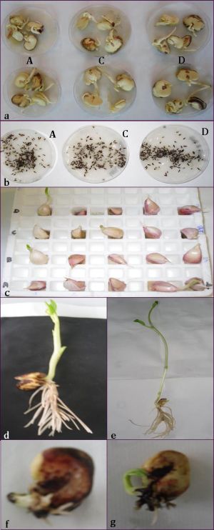 | Fig. 1- Germination of <i>V. faba</i> <b>(a)</b> and <i>L. sativa</i> <b>(b)</b> seeds, and <i>A. sativum</i> gloves <b>(c)</b> after 3 days of treatment by tap water (control) and textile effluent. Physiological aspect of <i>V. faba</i> shoots after 7 days of treatment by: <b>(d)</b> tap water (control); <b>(f)</b> textile effluent and after 9 days of treatment by: <b>(e)</b> tap water (control); <b>(g)</b> textile effluent. |
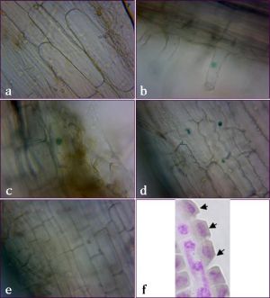 | Fig. 2- Direct Green BL dye diffusion in <i>L. sativa</i> L. root cells after one hour exposure: <b>(a)</b> control; <b>(b)</b> and <b>(c)</b> penetration by root hairs and surface accumulation in the epidermis at pH = 3.5; <b>(d)</b> deep accumulation in the inner parenchyma at pH = 4.5; <b>(e)</b> diffusion blockage of the dye through the cell membrane at pH = 9.5; <b>(f)</b> Global view of the cell layers isolated from the root tip region of <i>L. Sativa</i> L. in which the cytoplasm appeared dark colored due to dyes diffusion after 3 days of textile effluent exposure. |
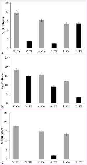 | Fig. 3- Mitotic index frequencies in <i>Vicia faba</i> L. (V), <i>Allium sativum</i> L. <b>(a)</b> and <i>Lactuca sativa</i> L. (L) after the treatment of root tip apices with textile effluent (TE) for 3 days <b>(a)</b> (primary roots), 7 <b>(b)</b> and 9 days <b>(c)</b> (secondary roots). In each experiment, the values of MI were compared to the control (Ctr) grown in pure water. The results are expressed as means ± s.d. (bar) of five different cytological analyses (1000 nuclei analyzed per slide). |
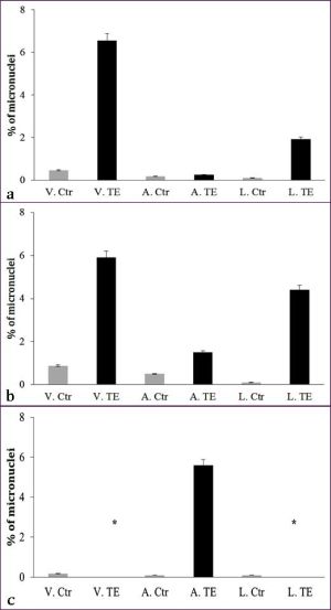 | Fig. 4- Micronuclei frequencies in <i>Vicia faba</i> L. (V), <i>Allium sativum</i> L. <b>(a)</b> and <i>Lactuca sativa</i> L. (L) after the treatment of root tip apices with textile effluent (TE) for 3 days <b>(a)</b> (primary roots), 7 <b>(b)</b> and 9 days <b>(c)</b> (secondary roots). In each experiment, the values of micronuclei were compared to the control (Ctr) grown in pure water. The asterisk means that all cells nuclei are fragmented. The results are expressed as means ± s.d. (bar) of five different cytological analyses (1000 nuclei analyzed per slide). |
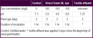 | Table 1- Experimental conditions of Direct Green BL dye diffusion in L. sativa L. root cells. |
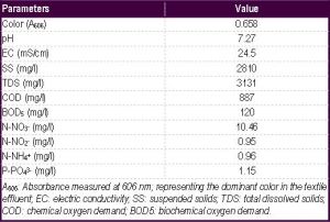 | Table 2- Physico-chemical characterization of real textile effluent |
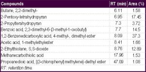 | Table 3- Main organic compounds identified in textile effluent using GC/MS |
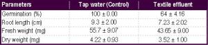 | Table 4- Effect of textile effluent on growth parameters of 15 days L. sativa shoots (n = 3, mean ± s.d.). |