


ISSN : 0976-8769
EISSN : 0976-8777



SHINOHARA H.1, NOGUCHI S.2, KUMAZAKI M.3, YAMADA N.4, ITO T.5, OYAMA M.6, ITO Y.7, OTSUKI Y.8, NAITO S.9, IINUMA M.10, AKAO Y.11*
1Gifu Pharmaceutical University, 1-25-4 Daigaku-nishi, Gifu 501-1196, Japan.
2United Graduate School of Drug Discovery and Medical Information Sciences, Gifu University, 1-1 Yanagido, Gifu 501-1193, Japan.
3Department of Biomolecular Science, Faculty of Engineering, Gifu University, 1-1 Yanagido, Gifu 501-1193, Japan.
4United Graduate School of Drug Discovery and Medical Information Sciences, Gifu University, 1-1 Yanagido, Gifu 501-1193, Japan.
5Gifu Pharmaceutical University, 1-25-4 Daigaku-nishi, Gifu 501-1196, Japan.
6Gifu Pharmaceutical University, 1-25-4 Daigaku-nishi, Gifu 501-1196, Japan.
7Osaka Medical College, 2-7 Daigaku-cho, Takatsuki, Osaka 596-8686, Japan.
8Osaka Medical College, 2-7 Daigaku-cho, Takatsuki, Osaka 596-8686, Japan.
9Department of Urology, Graduate School of Medical Sciences, Kyushu University, 6-10-1 Hakozaki, Higashi-ku, Fukuoka, Japan.
10Gifu Pharmaceutical University, 1-25-4 Daigaku-nishi, Gifu 501-1196, Japan.
11United Graduate School of Drug Discovery and Medical Information Sciences, Gifu University, 1-1 Yanagido, Gifu 501-1193, Japan.
* Corresponding Author : yakao@gifu-u.ac.jp
Received : 03-07-2013 Accepted : 10-07-2013 Published : 12-11-2013
Volume : 3 Issue : 1 Pages : 29 - 34
J Toxicol Res 3.1 (2013):29-34
10-Hydroxy-2-decenoic acid, which is a specific component of royal jelly, has anti-cancer effects. For the purpose of enhancing the anti-cancer activity of its medium-chain fatty acid derivatives, we synthesized 102 different derivatives and performed screening for their activity. Among them, several compounds exhibited a marked growth inhibition of K562 and NKB1 cells. Importantly, the apparent conversion from LC3B I to II and the down-regulation of E2F1 were found in both cell lines after treatment with AIC-47. Silencing E2F1 in both types of cells also induced autophagy. In vivo NKB1-cell/xenografted mice experiment indicated that the intraperitoneal injection of AIC-47 induced a significant anti-tumor effect through autophagic cell death in the 45 mg/kg-administered group, as observed in the in vitro experiment. The 3-decenoic acid derivative AIC-47 showed its anti-tumor effect by inducing autophagic cell death through down-regulation of E2F1 in both in vitro and in vivo models.
3-decenoic acid derivatives, anti-cancer agent, autophagic cell death, E2F1.
The components of royal jelly, a substance secreted by worker bees, have been well identified [1] . Several components have been reported to exhibit various functions allowing them to be classified as “functional†foods [2] . Among them, 10-hydroxy-2-decenoic acid is one of the specific components of royal jelly, and it is known to have an anti-cancer effect [3] . For the purpose of enhancing the anti-cancer activity of 10-hydroxy-2-decenoic acid, we synthesized 102 kinds of decenoic acid derivatives (AIC-1 to 102). In the screening of these derivatives, several 3-decenoic acid derivatives showed potent growth inhibition of human leukemia K562 cells.
Autophagy is a catabolic pathway for degradation and recycling of organelles and cytoplasmic contents to maintain cellular homeostasis. Autophagy can be induced by stress conditions such as starvation, mitochondrial dysfunction and infection. Although autophagy is considered as a survival response to various stressors, it is also involved in a particular type of cell death referred to as autophagic cell death [4,5] . Cell death is categorized into several types by morphology. Autophagic cell death is characterized by the sequestration of portions of the cytoplasm into autophagosomes, which appear as vacuoles in the cells. The machinery involved in this form of cell death and autophagy-related proteins are understood, but the signaling pathways linked with this machinery are not yet fully understood.
E2F1 regulates programmed cell death such as apoptosis or autophagy [6] . E2F1 induces apoptosis by up-regulating p53. E2F1 can also induce autophagy by up-regulating the expression of the autophagy genes LC3, Atg1, Atg5 and DRAM in response to DNA damage [7] . In cancer cells harboring neither wild p53 nor Rb, it is not known which stimulation and signaling pathways are associated with E2F1 activity.
In this study, we examined the mechanism of cell death induced by 3-decenoic acid derivatives. Our data indicate that 3-decenoic acid derivatives induced autophagic cell death in vitro and in vivo mainly through down-regulation of E2F1 and attenuation of the PI3K/Akt signal pathway.
Phosphatase Inhibitor Cocktails 2 and 3 were obtained from Sigma (St. Louis, MO, USA). Antibodies against the following human proteins were purchased from Cell Signaling Technology (MA, USA): Akt, phospho-Akt (Ser217/221), p44/42 MAP kinase (ERK), phospho-p44/42 MAPK (Thr202/Tyr204), cyclin D1, CDK6, c-Myc. Anti-mouse IgG HRP-linked antibody and anti-rabbit IgG HRP-linked antibody were also purchased from Cell Signaling Technology. Antibodies against human PARP-1 and human Bcl-2 were from Santa Cruz Biotechnology (CA, USA). The antibody against human Bax came from MBL (Nagoya, Japan). Monoclonal anti-β-Actin was obtained from Sigma.
Human leukemia cell line K562 was provided by HSRRB (Osaka, Japan). K562 cells were cultured in RPMI-1640 medium (Invitrogen, Carlsbad, CA, USA) supplemented with 10% (v/v) heat-inactivated FBS under an atmosphere of 95% air and 5% CO2 at 37°C. Cells of the human bladder cancer cell line NKB1 were a gift from the Cell Resource Center for Biomedical Research, Institute of Development, Aging, and Cancer of Tohoku University and were maintained according to the instruction provided. K562 cells overexpressing Bcl-2 were produced by transfecting K562 cells with the pIRESneo-Bcl-2 vector [8] . Each compound of the AIC-series was dissolved in dimethyl sulfoxide (DMSO) and added to the cell culture at a final concentration of DMSO (<0.3%) that showed no significant effect on the growth and differentiation of cells (data not shown). Viable cell numbers were measured by performing the Trypan-blue dye exclusion test.
For the morphological examination of cell death, K562 cells were stained with Hoechst 33342 (Calbiochem, San Diego, CA, USA), added to the culture medium at a concentration of 5 μg/ml. After incubation at 37°C for 30 min, the cells were collected, washed once with PBS, resuspended, pipetted dropwise onto a glass slide, and observed under a fluorescence microscope, Olympus CKX41N-FL (Olympus, Tokyo, Japan).
For preparation of cell lysate, cells were washed with phosphate-buffered saline (PBS) and harvested. The cell pellet was resuspended in lysis buffer comprising 10 mM Tris-HCl (pH 7.4), containing 0.1% SDS, 1% NP-40, 0.1% sodium deoxycholate, 150 mM NaCl, 1 mM EDTA, and 1% Phosphatase Inhibitor Cocktails and stood for 20 min on ice. After centrifugation at 13,000 rpm for 20 min at 4°C, the supernatants were collected as protein samples. Protein contents were measured with a DC Protein assay kit (Bio-Rad, Hercules, CA). Five micrograms of protein of each cell lysate was separated by SDS-PAGE by using an adequate percent of polyacrylamide gel and electroblotted onto a PVDF membrane (PerkinElmer Life Sciences, Waltham, MA). After blockage of nonspecific binding sites for 1 h by 5% nonfat milk in TBST (Tris-HCl buffer containing 0.1% Tween 20), the membrane was incubated overnight at 4°C with various primary antibodies. The membrane was then washed 4 times with TBST, incubated further with anti-mouse or anti-rabbit IgG HRP-linked antibody at room temperature, and then washed again 4 times with TBST. Proteins were detected by using an enhanced ECL kit (ParkinElmer) and chemiluminescence detector (LAS-4000, Fujifilm, Japan).
The cells treated or left untreated for 72 h with AIC-8, 47 or 82 (10 μM) and harvested and rinsed with PBS. The cells were then fixed for 8 h in 70% ethanol and rinsed again with PBS. After this rinse, the cells stained for 15 min at 37°C in propidium iodide (PI)/Triton X-100 staining solution containing RNase. Cell fluorescence was measured by use of a Tali Image-Based Cytometer (Invitrogen).
K562 cells treated or untreated with AIC-47 (10 μM) were harvested and rinsed with PBS. Cells were fixed for 2 h with 2% paraformaldehyde and 2.5% glutaraldehyde in 0.2 M phosphate buffer (pH 7.4, PB), rinsed in PB, and postfixed in 2% osmium tetraoxide for 2 h. After having been washed with PB, the cells were progressively dehydrated in a 10% graded series of 30-100% ethanol and then cleared in QY-1 (Nissin EM, Tokyo, Japan). Thereafter they were embedded in Epon 812 resin (TAAB Laboratories Equipment, Reading, UK), and thin sections (70 nm thickness) were prepared, after which they were stained with uranyl acetate and lead citrate and examined by transmission electron microscopy with a Hitachi-7650 (Hitachi, Tokyo, Japan), operating at 80 kV.
K562 and NKB1 cells for knockdown E2F1 were seeded into separate 6-well plates at a concentration of 0.5 × 10⁶ cells/well on the day before transfection. Short interfering RNA (siRNA) for E2F1 (10 nM) was used for transfection of the cells. Its sequence was 5’-UCGGCACCUGAGAAGCCUCUUGAAA-3’ (siR-E2F1; Invitrogen); and that of sequence of the non-specific control microRNA used (Hokkaido System Sciences, Sapporo Japan) was 5’-GGCCUUUCACUACUCCUCA-3’.
Animal experimental protocols were approved by Gifu University’s Committee for Ethics in Animal Experimentation, and animal experiments were conducted in accordance with the Guidelines for Animal Experiments of Gifu University. Human bladder cancer NKB1 cells were inoculated at 2 × 10⁶ cells/100 μl per site into the backs of athymic nude mice (day 0). The tumor-engrafted mice were sorted into 7 mice per group. AIC-47/ soybean oil emulsion was injected intraperitoneally once a week. The collection of the samples was performed on day 42, which was 1 week after the last injection. The levels of LC3B, Akt, and p-Akt in the samples were evaluated by Western blotting.
Each examination was performed in triplicate. Data are presented as means ± SD. Differences were statistically evaluated by one-way ANOVA followed by the t-test. The data for animal experiments were compared by Mann-Whitney U test. In all statistics data, the level of significance was considered to be p<0.05.
We examined the anti-cancer effect of 102 different 3-decenoic acid derivatives (AIC-1 to 102) on human leukemia K562 cells. Among them, approximately 50% of the AIC series of compounds exhibited a significant growth inhibition. Especially, several compounds that showed IC50 values of less than 10 μM, bore an amide or ester group [Fig-1] A and [Fig-1] B. The activities of AIC-8, -47, -49, and -82 were approximately the same as the activity of Etposide. Among them, AIC-8, -47, and -82 were selected for further experiments to elucidate the mechanism of their anti-cancer effects. Hoechst 33342 staining indicated that these compounds caused some of the treated cells to assume a typical apoptotic appearance such as chromatin condensation and fragmentation in some [Fig-2] A. However, no apparent biochemical finding of apoptosis was observed (e.g., PARP-1 cleavage: [Fig-2] B). We judged that apoptotic cell death occurred in only a few of the treated cells. Notably, however, the inactivation of PI3K/Akt signaling and down-regulation of Bcl-2 were significant [Fig-2] B. The MAPK signaling pathway including Erk1/2 was not activated [Fig-2] B.
(1A) Growth inhibition at 48 h after treatment of human leukemia K562 cells with 3-decenoic acid derivatives (AIC-8, -18, -47, -49, -82), 3-decenoic acid, Cytarabine or Etoposide. The cells were grown in the absence or presence of a 5 μM concentration of each compound. Data are expressed as the means ± SD of 3 different experiments. The cell viability of the control (DMSO alone) is indicated as 100%. The IC50 value of each compound is shown under the bar graph. p<0.05 is significant. (1B) Chemical structures of AIC-8, -18, -47, -49, -82.
(2A) Cells were stained by Hoechst 33342 (magnification ×100). White arrows indicate apoptotic cells. (2B) Expression and cleavage of PARP-1, and expression of Bcl-2 family proteins, MAP kinases and PI3K/Akt signaling molecules in K562 cells treated with AIC-8, -47 or -82 (5 μM & 10 μM) and examined by Western blot analysis.
Next, we analyzed the cell-cycle profile of K562 cells treated with these compounds, with the result being that the population of G0/G1 fraction was clearly increased at 72 h after the treatment with any of these 3 compound; in contrast, the population of cells in the G2/M phases was consistently decreased [Fig-3] A. The expression levels of cell cycle-related proteins, e.g., cyclin D1, CDK6, and c-myc, all of which function in the transition from the G1 to S phase, were significantly down-regulated [Fig-3] B. In order to examine whether the growth inhibition accompanied by the G0/G1 arrest was due to autophagy, we performed Western blot analysis of LC3B, a marker protein of autophagy. As shown in [Fig-4] A, a clear conversion from LC3B I to II was found in cells treated any of the 3 compounds tested. Unexpectedly, the expression level of Beclin-1 tended to be reduced slightly. Interestingly, the expression of E2F1 was markedly down-regulated in all of the treated cells [Fig-4] A. Furthermore, the electron-microscopic study demonstrated that autophagosomes, which contained multiple lipid-droplets, had appeared in the cells treated with AIC-47, which finding indicated that this 3-decenoic acid derivative had induced autophagy [Fig-4] B.
(3A) Cell number percentage in each phase are expressed as means ± SD of 3 different experiments. p<0.05 is significant. (3B) Expression of cyclin D1, CDK6, and c-myc after treatment of K562 cells with 5 μM AIC-8, -47 or -82 was examined by Western blot analysis.
Since there are several reports indicating that Bcl-2 is associated with both autophagy and apoptosis [9,10] , we produced a Bcl-2- overexpressing K562 cell line [Fig-5] . When Bcl-2 overexpressants were treated with AIC-47, the transition of LC3B I to II was not observed, as estimated by Western blot analysis. As a result, Bcl-2 overexpression did not rescue the cells from the AIC-47-induced autophagy. Next, we examined whether AIC-47 could induce autophagy in a cancer cell line aside from K562 leukemia, because we wanted to examine the in vivo anti-cancer effect of AIC-47 by using tumor-implanted mice. Finally, we found that AIC-47 induced a typical autophagy in human bladder NKB1 cells among the several cell lines tested. Also, AIC-47 effected a significant dose-dependent growth inhibition of these cells [Fig-6] A. Western blot analysis indicated the conversion of LC3B I to II, down-regulation of Bcl-2, and inactivation of PI3K/Akt signaling [Fig-6] B, as were observed in K562 cells [Fig-2] B and [Fig-4] A.
(4A) Conversion of LC3B I to II, and expression of Beclin-1 and E2F1 in AIC-8, -47 or -82 (5 μM and 10 μM)-treated K562 cells were examined by Western blot analysis. (4B) Morphology of the treated cells was examined by using TEM. K562 cells were treated with vehicle (control) or 10 μM AIC-47 for 72 h. White arrow indicates autophagosome/autolysosome containing a lipid droplet.
Expression and phosphorylation of PI3K/Akt signaling molecules, and expression of LC3B, Beclin-1, p27 and Sirt-1 in K562 cells or in Bcl-2-overexpressing K562 cells treated with AIC-47 (5 or 10 μM) and examined by Western blot analysis.
(6A) Growth inhibition of NKB1 cells at 72 h after the treatment with AIC-47. Data are expressed as means ± SD of 3 different experiments. p<0.05 is significant. (6B) Expression of LC3B, PI3K/Akt signaling molecules, Bcl-2, and E2F1 in AIC-47 (20 or 50 μM)-treated NKB1 cells was examined by Western blot analysis.
In order to elucidate the mechanism of AIC-47-induced autophagy, we focused on E2F1 down-regulation observed in both K562 and NKB1 cells after treatment with AIC-47. When we silenced E2F1 by using siRNA for E2F1, the conversion of LC3B I to II in both cell types was clearly observed at 72 h after their treatment with siRNA for E2F1 [Fig-7] A, [Fig-7] B. The combined treatment with siR-E2F1 and AIC-47 showed synergistic effects on the conversion of LC3B I to II.
The cells were transfected with siRNA for E2F1 (10 nM) in the presence of AIC-47 or without AIC-47 for 72 h. The expression of E2F1 and conversion of LC3B I to II were examined by Western blot analysis.
In order to confirm whether the AIC-47 induced autophagic cell survival or death, we pre-treated either type of cells with bafilomycin, a drug that interferes with the fusion of lysosomes to autophagosomes [11] , prior to treatment with AIC-47. Such pretreatment increased the number of viable cells compared with that for AIC-47-treated controls [Fig-8] A and [Fig-8] B. These findings taken together indicate that AIC-47 induced autophagic cell death, not survival, in both cell lines.
K562 and NKB1 cells were pre-incubated with 10 nM bafilomycin A1 and then co-treatment with AIC-47 for 72 h and then cell growth was examined. Data are expressed as the means ± SD of 3 different experiments. p<0.05 is significant.
Next, we performed an in vivo experiment to examine the anti-tumor effect of AIC-47. Human bladder NKB1 cells were inoculated subcutaneously into nude mice. As shown in [Fig-9] A, the intraperitoneal injection by AIC-47 (45 mg/kg) in an oil mixture once a week caused a significant suppression of tumor growth from day 21. In the case of 45 mg/kg, the tumor growth (< 50 mm3) was extremely suppressed from day 14 up to day 42. However, in the case of 9 mg/kg, the tumor was gradually increased with the same pattern as seen in the control group. Thus, these findings indicate that the anti-tumor suppressive activity of AIC-47 was probably dose dependent. We also estimated the transition of LC3B I to II in the tumor tissue samples by Western blotting [Fig-9] B. As a result, most of the tumor samples from the higher-dose group indicated a greater LC3B I to II transition than those in the control and lower-dose groups. On the other hand, Akt and Akt phosphorylation levels were slightly decreased in the higher-dose group compared with those in the control group, but remained almost the same in lower-dose group [Fig-9] B. Pathologically, onion-like regions that consisted of swollen cells with vacuoles were found in the higher-dose group (data not shown).
In the current study, we demonstrated that 3-decenoic acid derivatives functioned as inducers of autophagic cell death in both human leukemia K562 and bladder cancer NKB1 cells. The IC50 values obtained with K562 cells were almost the same as that value for Etoposide, AIC-47, one of the compounds bearing an amide group, exhibited one of the stronger anti-cancer activities among the 3-decenoic acid derivatives. Basically, the inactivation of PI3K/Akt signaling pathway and down-regulation of Bcl-2 and E2F1 were commonly found in response to treatment with various derivatives. Morphologically, we confirmed that the growth suppression by AIC-47 was due to autophagy. Interestingly, the autophagosomes in the treated cells contained lipid-droplets, indicating so-called lipophagy. Biochemically, the transition from LC3B I to II effected in both K562 and NKB1 cells by AIC-47 was clearly demonstrated, indicating autophagic cell death, as did a functional experiment using bafilomycin, an inhibitor of autophagy. However, Beclin-1 expression was almost unchanged after the treatment of either cell type with the derivatives.
(9A) NKB1 cells-xenografted mice were treated with AIC-47 (9 mg/kg or 45 mg/kg, IP) or an equal amount of soy bean oil as control once a week for 6 weeks. Tumor volumes were measured every week in each group. Data are expressed as the means ± SD. T = injection of tumor cells; S = time of sacrifice. p<0.05 is significant. (9B) Conversion of LC3B I to II and phosphorylation of Akt in the tumor samples on day 42 were examined by Western blot analysis.
We considered that AIC-47 perturbed not only the expression of glycolytic enzymes through the down-regulation of the PI3K/Akt signaling pathway, but also enzymes required for β oxidation of fatty acids; because AIC-47 induced lipophagy, and fatty acid analysis indicated that the level of C13 myristic acid was increased among the metabolites of fatty acids in the AIC-47-treated cells (data not shown). It is possible that AIC-47 disturbed the entry of fatty acid metabolites into the mitochondrial inner membrane for β oxidation. Therefore, we suspect that Bcl-2, which is located in the inner membrane, might be associated with the translocase needed for the entry of fatty acids for β oxidation. However, the overexpression of Bcl-2 could neither rescue the cells from autophagic cell death nor affect the PI3K/Akt signaling, which findings may also reflect the unchanged expression level of Beclin-1.
Tumor-suppressor proteins including RB and p53, positively regulate autophagy [6,12,13] , as do upstream regulators of RB, including the cyclin-dependent kinase inhibitors p16INK4a and p27kip1 [14,15] . E2F1, a pro-apoptotic member of the E2F family, is a master gatekeeper for cell-cycle progression [16] . The RB-E2F1 axis has an important role in the regulation of autophagy [6,7] . In our study, RB and p16INK4a proteins were not expressed at detectable levels in either K562 or NKB1 cells (data not shown). Therefore, AIC-47-triggered down-regulation of E2F1 and the silencing of E2F1 with siR-E2F1 in both cells induced autophagy, indicating that the inhibition of E2F1 activity could be in part responsible for the triggering of autophagy in AIC-47-induced autophagy. Both PI3K/Akt signaling and E2F1 inactivation could cause the autophagic cell death, not survival. However, it remains unknown as to how AIC-47 effected the down-regulation of E2F1. The fact that autophagy is one of the biological phenotypes for cell survival may have important overtones for the therapy of cancer. In fact, autophagy may be closely associated with chemo- and radio-resistance [17,18] .
Among the 102 different medium-chain fatty acid derivatives examined, which ranged from C8 to C12, several kinds of 3-decenoic acid derivatives exhibited a potent anti-cancer activity with IC50 values ranging from 2.1 to 5.2 μM. It has been reported that the long-chain fatty acid palmitic acid induces autophagy at 0.25 mM through lipotoxicity, which is independent of mTOR regulation [19] . This event is due to excess fat accumulation. AIC-47 showed no growth inhibitory effect at the concentrations used in this study for normal human lymphocytes stimulated with Concanavalin A (data not shown). Based on the results from our experiments, the 3-decenoic acid derivatives functioned as signaling molecules. Recently, many orphan G-protein-coupled receptors (GPCR) have been found; and a GPCR deorphanizing strategy successfully identified multiple receptors for free fatty acids [20] . Free fatty acids have been proven to act as signaling molecules; and GPR40 and GPR120, which are receptors for these free fatty acids, are activated by medium- to long-chain free fatty acids [20,21] . However, both receptors are associated with cell proliferation and inhibition of apoptosis when treated with free fatty acids [20,21] . Especially, linolenic acid promotes the activation of ERK and PI3K/Akt pathways mainly via GPR120, leading to an anti-apoptotic effect [20,22] . In our experiment using some long-chain fatty acid derivatives, apoptotic cell death was induced by their compounds. It is not known whether the metabolites from 3-decenoic acid derivatives or the derivatives per se specifically affect signaling molecules or gene expression of transcription factors through the association with CD36 and the receptors GPR40 and GPR120 [23] . Further study will be needed to disclose the detailed mechanism of the function of 3-decenoic acid derivatives in autophagic cell death.
The 3-decenoic acid derivatives showed anti-cancer effect by inducing autophagic cell death through down-regulation of E2F1. These compounds showed anti-tumor effect both in vitro and in vivo.
None declared.
This work was supported by a grant-in-aid for scientific research from the Ministry of Education, Science, Sports, and Culture of Japan.
[1] Weaver N., Johnston N.C., Benjamin R. and Law J.H. (1968) Lipids, 3, 535-538.
» CrossRef » Google Scholar » PubMed » DOAJ » CAS » Scopus
[2] Viuda-Martos M., Ruiz-Navajas Y., Fernandez-Lopez J. and Perez-Alvarez J.A. (2008) J. Food Sci., 73, R117-124.
» CrossRef » Google Scholar » PubMed » DOAJ » CAS » Scopus
[3] Townsend G.F., Brown W.H., Felauer E.E. and Hazlett B. (1961) Can. J. Biochem. Physiol., 39, 1765-1770.
» CrossRef » Google Scholar » PubMed » DOAJ » CAS » Scopus
[4] Baehrecke E.H. (2005) Nat. Rev. Mol. Cell. Biol., 6, 505-510.
» CrossRef » Google Scholar » PubMed » DOAJ » CAS » Scopus
[5] Codogno P. and Meijer A.J. (2005) Cell Death Differ., 12(2), 1509-1518.
» CrossRef » Google Scholar » PubMed » DOAJ » CAS » Scopus
[6] Jiang H., Martin V., Gomez-Manzano C., Johnson D.G., Alonso M., White E., Xu J., McDonnell T.J., Shinojima N. and Fueyo J. (2010) Cancer Res., 70, 7882-7893.
» CrossRef » Google Scholar » PubMed » DOAJ » CAS » Scopus
[7] Polager S., Ofir M. and Ginsberg D. (2008) Oncogene, 27, 4860-4864.
» CrossRef » Google Scholar » PubMed » DOAJ » CAS » Scopus
[8] Akao Y., Maruyama W., Shimizu S., Yi H., Nakagawa Y., Shamoto-Nagai M., Youdim M.B., Tsujimoto Y. and Naoi M. (2002) J. Neurochem., 82, 913-923.
» CrossRef » Google Scholar » PubMed » DOAJ » CAS » Scopus
[9] Drullion C., Tregoat C., Lagarde V., Tan S., Gioia R., Priault M., Djavaheri-Mergny M., Brisson A., Auberger P., Mahon F.X. and Pasquet J.M. (2012) Cell Death Dis., 3, e373.
» CrossRef » Google Scholar » PubMed » DOAJ » CAS » Scopus
[10] Shimizu S., Kanaseki T., Mizushima N., Mizuta T., Arakawa-Kobayashi S., Thompson C.B. and Tsujimoto Y. (2004) Nat. Cell Biol., 6, 1221-1228.
» CrossRef » Google Scholar » PubMed » DOAJ » CAS » Scopus
[11] Yamamoto A., Tagawa Y., Yoshimori T., Moriyama Y., Masaki R. and Tashiro Y. (1998) Cell Struct. Funct., 23, 33-42.
» CrossRef » Google Scholar » PubMed » DOAJ » CAS » Scopus
[12] Crighton D., Wilkinson S. and Ryan K.M. (2007) Autophagy, 3, 72-74.
» CrossRef » Google Scholar » PubMed » DOAJ » CAS » Scopus
[13] Ciavarra G. and Zacksenhaus E. (2011) Autophagy, 7, 544-546.
» CrossRef » Google Scholar » PubMed » DOAJ » CAS » Scopus
[14] Komata T., Kanzawa T., Takeuchi H., Germano I.M., Schreiber M., Kondo Y. and Kondo S. (2003) Br. J. Cancer, 88, 1277-1280.
» CrossRef » Google Scholar » PubMed » DOAJ » CAS » Scopus
[15] Naqsh e Zahra S., Khattak N.A. and Mir A. (2013) Theor. Biol. Med. Model, 10, 1.
» CrossRef » Google Scholar » PubMed » DOAJ » CAS » Scopus
[16] Johnson D.G., Schwarz J.K., Cress W.D. and Nevins J.R. (1993) Nature, 365, 349-352.
» CrossRef » Google Scholar » PubMed » DOAJ » CAS » Scopus
[17] Carew J.S, Nawrocki S.T. and Cleveland J.L. (2007) Autophagy, 3, 464-467.
» CrossRef » Google Scholar » PubMed » DOAJ » CAS » Scopus
[18] Zhuang W., Qin Z. and Liang Z. (2009) Acta Biochim. Biophys. Sin. (Shanghai), 41, 341-351.
» CrossRef » Google Scholar » PubMed » DOAJ » CAS » Scopus
[19] Tan S.H., Shui G., Zhou J., Li J.J., Bay B.H., Wenk M.R. and Shen H.M. (2012) J. Biol. Chem., 287, 14364-14376.
» CrossRef » Google Scholar » PubMed » DOAJ » CAS » Scopus
[20] Katsuma S., Hatae N., Yano T., Ruike Y., Kimura M., Hirasawa A. and Tsujimoto G. (2005) J. Biol. Chem., 280, 19507-19515.
» CrossRef » Google Scholar » PubMed » DOAJ » CAS » Scopus
[21] Wauquier F., Philippe C., Leotoing L., Mercier S., Davicco M.J., Lebecque P., Guicheux J., Pilet P., Miot-Noirault E., Poitout V., Alquier T., Coxam V. and Wittrant Y. (2013) J. Biol. Chem., 288, 6542-6551.
» CrossRef » Google Scholar » PubMed » DOAJ » CAS » Scopus
[22] Soto-Guzman A., Robledo T., Lopez-Perez M. and Salazar E.P.(2008) Mol. Cell Endocrinol., 294, 81-91.
» CrossRef » Google Scholar » PubMed » DOAJ » CAS » Scopus
[23] Koonen D.P., Glatz J.F., Bonen A. and Luiken J.J. (2005) Biochim. Biophys. Acta, 1736, 163-180.
» CrossRef » Google Scholar » PubMed » DOAJ » CAS » Scopus
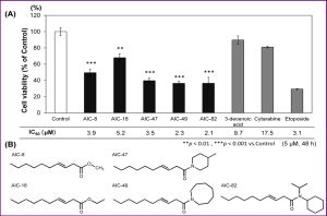 | Fig. 1- 3-Decenoic acid derivatives induced growth inhibition in K562 cells. |
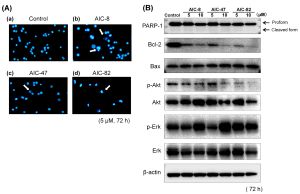 | Fig. 2- Expression of the molecules related to cell growth and apoptotic signaling pathway treated with 3-decenoic acid derivatives in K562 cells. |
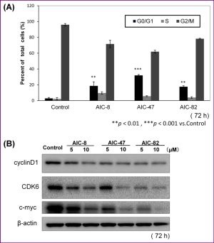 | Fig. 3- 3-decenoic acid derivatives arrested cell cycle in G0/G1 Phase. |
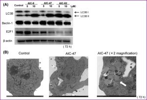 | Fig. 4- Induction of autophagy after treatment of K562 cells with 3-decenoic acid derivatives, as judged by Western blotting and electron microscopic analyses. |
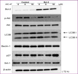 | Fig. 5- The effect of overexpression of Bcl-2 on the expression of growth and autophagy related proteins in K562 cells. |
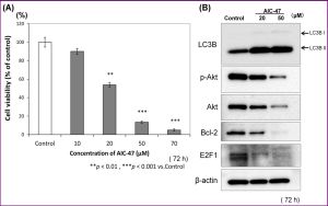 | Fig. 6- Induction of autophagy in human bladder cancer NKB1 cells by AIC-47. |
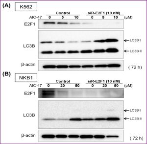 | Fig. 7- Induction of autophagy in K562 (7A) and NKB1 (7B) cells by silencing E2F1. |
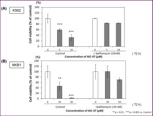 | Fig. 8- Inhibition of autophagy by bafilomycin A1 in K562 (8A) and NKB1 (8B) cells. |
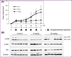 | Fig. 9- Anti-tumor effect of AIC-47 on mice xenografted with human NKB1 cells. |