


ISSN : 0976-7061
EISSN : 0976-707X



SABNIS A.S.1*
1Department of Anatomy, K.J. Somaiya Medical College, Sion, Mumbai- 400 022, MS, India.
* Corresponding Author : dranjus2003@yahoo.com
Received : 10-04-2013 Accepted : 25-04-2013 Published : 05-10-2013
Volume : 4 Issue : 1 Pages : 54 - 56
J Clin Res Lett 4.1 (2013):54-56
DOI : http://dx.doi.org/10.9735/0976-7061.4.1.54-56
Conflict of Interest : None declared
Background- Profunda femoris artery (PFA) is the largest branch of femoral artery which supplies all the muscles of thigh. During the procedures like arterial catheterization damage to PFA may invite unwanted and unexpected complications. Precise anatomy of PFA forms strong foundation to minimize complications. Materials and Methods- Femoral triangles of 60 lower limbs of 27 male and 3 female embalmed cadavers were dissected to study origin, course and branches of PFA. The external diameter of femoral artery and PFA was measured. Results- In all the lower limbs PFA was present and in majority of cases the commonest site of origin was posterolateral aspect of femoral artery. The mean distance from inguinal ligament from where PFA was arising from femoral artery was 3.2 cm. In majority of lower limbs lateral circumflex femoral artery was arising from PFA. Conclusion- Point of origin of PFA was similar in some cases and different in other cases. Detail knowledge regarding variations in point of origin, branches of PFA is clinically significant and should not be ignored.
Profunda femoris artery (PFA), 60 lower limbs
PFA and its variations are the topics of interest for surgeons and anatomists. Along with femoral artery, PFA is also the important artery of anterior aspect of thigh. PFA is the branch of femoral artery which arises from lateral aspect of femoral artery 3.5 cm distal to inguinal ligament [1] . It gives lateral and medial circumflex femoral arteries from lateral and medial aspect respectively [2] . PFA shows variations in terms of point of origin, course and branches. These variations have received attention of surgeons, radiologists, cardiologists and anatomists.
The femoral artery is second site of choice after radial artery for placement of an arterial line. When easily accessible veins are collapsed femoral vein is used for collection of blood. Thus femoral triangle is clinically useful and important area for accessing vessels. Origin of PFA from femoral artery at short distance distal to inguinal ligament. Shorter the distance of origin of PFA higher is the risk of iatrogenic damage of PFA. Any variation in PFA should be taken in account to prevent unexpected and unpleasant complications. So we studied PFA in detail and any variations came across were recorded.
Femoral triangles of 60 lower limbs of 27 male and 3 female embalmed cadavers obtained from department of anatomy were dissected. to study origin, course and branches of PFA. After removing skin and superficial fascia, femoral artery, vein and nerve was identified. PFA was searched on the lateral aspect of femoral artery. The origin, course and branches were noted. The distance of origin of PFA from midpoint of inguinal ligament was measured with the help of scale. The width of femoral and PFA was measured with the help of divider and scale.
1. In all lower limbs PFA was present.
2. In 52 and 8 lower limbs PFA was originating from lateral [Fig-1] , [Fig-3] and [Fig-5] and posterolateral aspect [Fig-4] of femoral artery respectively.
3. The point of origin was similar on both sides in 46 lower limbs [Fig-5] while it was different on two sides in 14 lower limbs (6 males and one female) [Fig-1] .
4. The mean distance from inguinal ligament from where PFA was arising from femoral artery was 3.2 cm [Table-1] . While in 18 lower limbs PFA was arising just below the inguinal ligament [Fig-2] , [Fig-3] .
5. The mean width of femoral artery was 10 cm and of PFA was 6 mm.
6. Absence of lateral circumflex femoral artery was seen in 2 lower limbs. Ascending, transverse and descending branches were arising from PFA [Fig-2] .
7. In 10 lower limbs lateral circumflex femoral artery was originating from femoral artery [Fig-4] .
Procedures like arteriography, Doppler imaging, vascular reconstruction of proximal leg necessitates precise anatomy of PFA along with the femoral artery. Variations of PFA and their awareness will definitely reduce the risk of damaging them.
Origin of PFA is from lateral aspect of femoral artery [1] , in 46% it is from posterior aspect, in 30% from posterolateral aspect and in 23% is from lateral aspect [3] . In the present study 86% of lower limbs showed origin of PFA from lateral aspect of femoral artery [Fig-1] , [Fig-3] and [Fig-5] and in 14% of lower limbs it was from posterolateral aspect [Fig-4] . The distance between midpoint of inguinal ligament and origin of PFA is clinically important. Too much proximal origin and or too much distal origin of PFA may invite unwanted problems. Judkin technique is undertaken where femoral artery is approached by puncturing the vessel 1 to 3 cm below the inguinal ligament [4] . Knowledge of the site of origin of PFA helps in avoiding iatrogenic femoral A-V fistulas or severe secondary hemorrhage when performing a femoral arterial puncture [2] . Following table shows average distance from midpoint of inguinal ligament measured by various authors.
In 1 out of 431 cases PFA was originated above the inguinal ligament, in 1.6% it was originated deep to inguinal ligament and in 3.01% cases it was originated half inch below the inguinal ligament [12] . In the present study in 18 lower limbs PFA was arising just below the inguinal ligament [Fig-2] , [Fig-3] . The external and internal diameters are important as it gives judgment of the diameter of guide wire which is to be used in procedures. We have measured the width of the femoral and PFA with the help of divider and scale. The mean width of femoral artery was 10 cm and of PFA was 6 mm. In two lower limbs there was no much difference in between caliber of femoral and PFA [Fig-3] . PFA acts as a collateral vessel in the occlusion of the femoral artery and for this important function it has to have a larger caliber [13] . In majority of cases (40) where the point of origin of PFA was similar on both sides, same width was found [Fig-5] while in remaining 6 lower limbs width was different on both sides. Out of 14 lower limbs where the point of origin of PFA was different on two sides width was found same in 10 lower limbs and different in 4 lower limbs [Fig-1] .
PFA gives lateral and medial circumflex femoral artery in addition to perforating branches. Lateral circumflex femoral artery gives ascending, transverse and descending branches. Variations are seen in branching pattern of PFA unilaterally and or bilaterally.
Following table shows variations noticed by various authors.
The knowledge of variations in the height origin of PFA and its branches distribution is of great significance for preventing flap necrosis particularly tensor fascia latae, when used in plastic and reconstructive surgery [6] . Anatomical facts should be considered before planning different diagnostic and therapeutic interventions on femoral artery and its branches [7] .
Details of origin and branches of femoral and PFA can be studied with high resolution ultrasonic imaging before any invasive procedure in the femoral triangle to achieve impressive goals.
[1] Standring S. (2004) Grays Anatomy, 40th ed., Philadelphia, Elsevier Churchill Livingstone, 1379-1380.
» CrossRef » Google Scholar » PubMed » DOAJ » CAS » Scopus
[2] Mc Minn R.M.H. (1994) Last’s Anatomy, 9th ed., London, Churchill Livingstone, 155-6.
» CrossRef » Google Scholar » PubMed » DOAJ » CAS » Scopus
[3] Samarawickrama M.B., Nanayakkara B.G., Wimalagunarathna K.W.R., Nishantha D.G., Walawage U.B. (2009) Galle Medical Journal, 14(1), 31-34.
» CrossRef » Google Scholar » PubMed » DOAJ » CAS » Scopus
[4] Baptist M., Sultana F., Hussain T. (2007) Professional Med. J., 14(3), 523-527.
» CrossRef » Google Scholar » PubMed » DOAJ » CAS » Scopus
[5] Snell R.S. (1992) Clinical Anatomy of Medical Students, 4th ed., Little Brown and Co, Boston, 607.
» CrossRef » Google Scholar » PubMed » DOAJ » CAS » Scopus
[6] Vuksanovic B.A., Stefanivic N., Pavlovic S., Duraskosvic R., Randelovic J. (2007) Facta Universitatis, Medicine and Biology, 14(3), 112-116.
» CrossRef » Google Scholar » PubMed » DOAJ » CAS » Scopus
[7] Prakash Jyoti K., Bhardwaj A.K., Jose B.A., Yadav S.K., Singh G. (2010) Rom. J. Morphol. Embryol., 51(1), 167-170.
» CrossRef » Google Scholar » PubMed » DOAJ » CAS » Scopus
[8] Dixit D., Kubavat D., Rathod S., Patel M., Singel T. (2011) Int. J. Biol. Med. Res., 2(4), 1084-1089.
» CrossRef » Google Scholar » PubMed » DOAJ » CAS » Scopus
[9] Bannister L.H., Berry M.M., Collins P. (1995) Gray’s Anatomy, 38th ed., Churchill livingstone, Medical division of Longman Group, UK, 1566-8.
» CrossRef » Google Scholar » PubMed » DOAJ » CAS » Scopus
[10] Siddharth P., Smith N.L., Mason R.A., Giron F. (1985) Anat. Rec., 212(2), 206-209.
» CrossRef » Google Scholar » PubMed » DOAJ » CAS » Scopus
[11] Chleborad W.P., Dawson D.L. (1990) Clinical Anatomy, 3(1), 33-40.
» CrossRef » Google Scholar » PubMed » DOAJ » CAS » Scopus
[12] Quain R. (1844) Anatomy of Arteries of the Human Body, London, Taylor and Walton, 476-8.
» CrossRef » Google Scholar » PubMed » DOAJ » CAS » Scopus
[13] Vaas F. (1975) Arch Chir. Neerl., 27(1), 25-34.
» CrossRef » Google Scholar » PubMed » DOAJ » CAS » Scopus
[14] Uzel M., Tanyeli E., Yildirim M. (2008) Folia Morphol., 67(4), 226-230.
» CrossRef » Google Scholar » PubMed » DOAJ » CAS » Scopus
[15] Fukuda H., Ashida M., Ishii R., Abe S., Ibukuro K. (2005) Surg. Radiol. Anat., 27(3), 260-264.
» CrossRef » Google Scholar » PubMed » DOAJ » CAS » Scopus
[16] Choi S.W, Park J.Y., Hur M.S., Park H.D., Kang H.J., HU K.S., Kim H.J. (2007) J. Craniofac. Surg., 18(4), 866-871.
» CrossRef » Google Scholar » PubMed » DOAJ » CAS » Scopus
[17] Tansatit T., Wanidchaploi S., Sanguansit P. (2008) J. Med. Assoc. Thai, 91(9), 1404-1409.
» CrossRef » Google Scholar » PubMed » DOAJ » CAS » Scopus
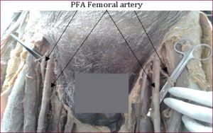 | Fig. 1- shows different point of origin and different caliber of PFA on two sides |
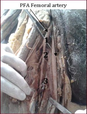 | Fig. 2- shows unilateral origin of PFA just below inguinal ligament and absence of lateral circumflex femoral artery. 1-ascending, 2-transverse and 3-descending branches are arising from PFA |
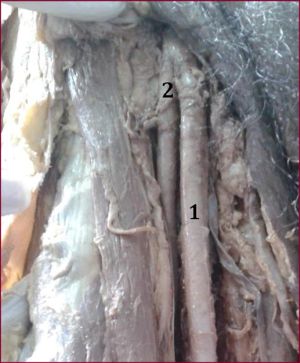 | Fig. 3- shows unilateral higher origin of PFA just below inguinal ligament from femoral artery on right side |
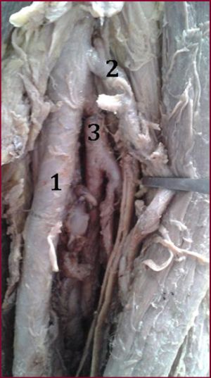 | Fig. 4- shows origin of lateral circumflex femoral artery (2) from femoral artery (1) directly and PFA (3) originated from posterolateral aspect of femoral artery |
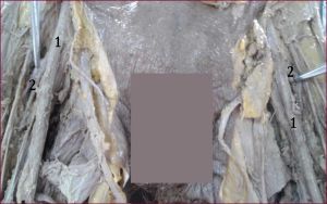 | Fig. 5- shows similar point of origin of PFA (2) from femoral artery (1) on both sides |
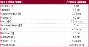 | Table-1 shows average distance of origin of PFA from midpoint of inguinal ligament |