


ISSN : 0976-8769
EISSN : 0976-8777



NORAZLINA M.1*, NORZALYANA N.M.Z.2, IMA-NIRWANA S.3
1Department of Pharmacology, Universiti Kebangsaan Malaysia, Medical Centre, Kuala Lumpur, Malaysia.
2Department of Biomedical Sciences, Faculty of Health Sciences, Universiti Kebangsaan Malaysia, Kuala Lumpur, Malaysia.
3Department of Pharmacology, Universiti Kebangsaan Malaysia, Medical Centre, Kuala Lumpur, Malaysia.
* Corresponding Author : azlina@medic.ukm.my
Received : 07-03-2013 Accepted : 11-04-2013 Published : 04-11-2013
Volume : 3 Issue : 1 Pages : 25 - 28
J Toxicol Res 3.1 (2013):25-28
Background- Palm vitamin E had been studied extensively for health and medicinal purposes. However, its toxic effects need to be elucidated. This study was done to determine the toxic effects of palm vitamin E in various dosages on the reproductive system of mice by determining the body weight, amount and size of pups delivered and post-mortem examination of the dams. Methods: This study was divided into two sub-studies i.e. giving palm vitamin E before and during pregnancy. A total of 56 mice were used in this study. The mice were divided into four groups; the control group, and groups supplemented with either palm vitamin E at 200mg/kg, 500mg/kg or 1000mg/kg. Supplementation of palm vitamin E before pregnancy involved giving palm vitamin E daily two weeks before pregnancy and no palm vitamin E given until birth. Supplementation of palm vitamin E during pregnancy involved giving palm vitamin E daily from the first day of pregnancy until birth, which was about 21 days. Body weights were recorded weekly. After delivery, the amount, weight and body length of pups were determined after which the dams were killed for post-mortem analysis of the uterus. Results: The results showed no significance difference (p>0.05) in the weight, amount and size of pups delivered amongst the groups for both sub-studies. Conclusion: Palm vitamin E at the dosages of 200 mg/kg, 500 mg/kg and 1000 mg/kg which was given before and during pregnancy did not cause any toxic effects on the reproductive system of mice.
female mice, palm vitamin E, reproductive, toxicity.
Vitamin E exists in two forms, tocotrienols and tocopherols. Both act as antioxidants and free radical scavengers. Recently, extensive studies had been carried out on tocotrienols and it was shown to exert beneficial effects in various conditions and diseases. It demonstrated hypocholesterolemic, anti-cancer and neuroprotective properties [1] . As an anti-cancer agent, tocotrienols had been shown to target multiple genes [2] . Tocotrienols had also been shown to have anti-osteoporotic effects [3,4] . One source of tocotrienols is the palm oil. Crude palm oil consists of 36% α-tocopherol, 31% γ-tocotrienol, 22% α-tocotrienol and 7% d-tocotrienol [5] . The health benefits of palm vitamin E may be attributed to the tocotrienol content.
In order for palm vitamin E to be used in clinical settings, its toxicological profile must be established. To date, not many toxicity studies have been done on tocotrienols or palm vitamin E as compared to tocopherols. Toxicity studies carried out on mice and rats which were given oral supplementation of palm vitamin E at the dose of 250, 500, 1000 and 2500 mg/kg for 30 days showed no adverse effects on the animals [6] . Non-observable-adverse-effects level (NOAEL) for tocotrienol is 0.19% (120 mg/kg body weight/day for male rats and 130 mg/kg body weight/day for female rats) when given in the diet for 13 weeks [7] . It was also reported that high palm oil levels in the diet induced liver toxicity [8] . In a recent study, palm vitamin E at the dose of 200, 500 and 1000 mg/kg was given to mice for either 14 days or 42 days. It was observed that palm vitamin E at 500 mg/kg may cause bleeding tendencies and renal impairment [9] .
In terms of effects on the reproductive system, α-tocopherol had been suggested to have a role as a therapeutic agent in pre-eclampsia [10] . Vitamin E, namely tocopherols, did not cause any toxicity on the reproductive system [11,12] . Very limited studies were done with tocotrienols. Tocotrienols given to pregnant rats were bioavailable to the fetal brain which suggests its role in gene expression profile of the developing fetal brain [13] . Considering the efficient transport of tocotrienols via the placenta and into the fetal brain, the study of its toxicity before and during pregnancy is justified.
In this study, we determined the toxic effects of palm vitamin E at various dosages given to mice before and during pregnancy. The data obtained will shed some light on the toxic effects of palm vitamin E and will be useful in future clinical applications using palm vitamin E.
Fifty-six female mice aged 1-2 months weighing 20-30 grams were obtained from the University’s Laboratory Animals Resource Unit. Sixteen male mice were also obtained and used for mating. The female mice were divided into two sub-studies i.e.
supplementation of palm vitamin E before pregnancy and
supplementation of palm vitamin E during pregnancy.
For both studies, the mice were randomly divided into 4 groups; control group (C) and groups supplemented with either palm vitamin E 200 mg/kg (PVE200), 500 mg/kg (PVE500) or 1000 mg/kg (PVE1000). The treatment was carried out as follows:
Sub-Study-i: The mice received palm vitamin E via oral gavage for 14 days before mating
Sub-study-ii: the mice received palm vitamin E via oral gavage from the first day of pregnancy until delivery.
The protocol was carried out based on the guidelines outlined by OECD [14] . Palm vitamin E was a gift from the Malaysian Palm Oil Board (Bangi, Selangor). It consisted of 24.82% α-tocopherol, 26.68 γ-tocotrienol, 20.73% α-tocotrienol and 13.32% δ-tocotrienol. A total of 1 g, 2.5 g and 5 g of palm vitamin E was dissolved in 10 ml olive oil to obtain the concentration of 200, 500 and 1000 mg/kg respectively. A volume of 0.02 ml for every 10 g of body weight was given via oral gavage. Supplementation was given once daily. The mice were kept individually in cages and were fed rat chow (Gold Coin, Port Klang, Selangor) and given tap water ad libitum.
The female and the male mice were placed in a cage for at least 24 hours up to a maximum of 3 days. The ratio of female to male in the cage was 4:1. To determine successful conception, a vaginal swab was carried out on the female mice every morning. The sample obtained from the swab was smeared on a glass slide. The slide was then dried and dipped into 95% alcohol for 1-2 minutes. It was then dipped into Schorr solution and rinsed with 75% alcohol for 1-2 minutes to remove excess colour. Once the slide was dried, sperm detection was made under light microscope with x100 magnification. Sperms were stained blue due to the Schorr solution.
The body weights of the female mice were recorded weekly. Within 24 hours of delivery, the number of pups as well as its body weight and body length were recorded. The dams were sacrificed within 24 hours of delivery and the uterine horns were exteriorized after opening the abdomen by midline incision. The sacs were inspected for sites of fetal resorption.
The study was carried out according to the guidelines of Universiti Kebangsaan Malaysia Animal Ethics Committee with the approval number FAR/2006/NORAZLINA/22-AUGUST/184-MAY-2007.
Results were presented as mean ± standard error of the mean (SEM). All data were analysed using the Statistical Package for Social Sciences software (SPSS 19.0). The Kolmogorov-Smirnov test was used for normality testing. ANOVA followed by Tukey’s HSD tests were used for normally distributed data while Kruskal-Wallis and Mann-Whitney tests were used for not normally distributed data.
[Fig-1] shows the body weight of mice supplemented with palm vitamin E before the pregnancy. There was no significant difference between the different groups. However, mice supplemented with 1000 mg/kg palm vitamin E during the pregnancy was significantly heavier compared to PVE200 group at week 3 [Fig-2] .
No significant difference was observed in all parameters in the different treatment groups for both sub-studies i.e. supplementation of palm vitamin E before pregnancy [Table-1] and supplementation of palm vitamin E during pregnancy [Table-2] .
No fetal resorption was observed except for 1 mouse in the 200 mg/kg group which received the supplementation before pregnancy. There were 2 fetal resorptions observed.
The role of vitamin E on reproductive system has been reported in several studies. Low levels of serum α-tocopherol were associated with abnormal semen pattern in men and anovulation in women [15] . In another study, low levels of α-tocopherol in seminal fluid were associated with low fertility in male subjects [16] . Alpha-tocotrienol was found to restore fertility in tocopherol-transport-protein deficient mice [17] . The role of vitamin E in the reproductive system is attributed to its antioxidant property. Oxidative stress influenced the function of the reproductive system and was associated with various abnormalities and pathological conditions involving the reproductive system [18] . Thus, antioxidants such as vitamin E are able to prevent the damage caused by oxidative stress.
Although vitamin E was found to have beneficial effects on the reproductive system, the effects of high doses of vitamin E on the reproductive system need to be elucidated. Thus, we investigated the effects of three different doses of palm vitamin E supplementation either before or during pregnancy on the reproductive system of female mice.
Body weights of the mice were not significantly different to each other when palm vitamin E was supplemented before pregnancy however mice given 1000 mg/kg of palm vitamin E during pregnancy had a significant increase in body weight compared to PVE200 group. The difference was observed at week 3. A previous study showed an association between vitamin E (α-tocopherol) and leptin in which vitamin E was negatively correlated with leptin in obese and non-insulin-dependent diabetic subjects [19] . Leptin is a hormone which acts on the brain to regulate food intake and energy expenditure. Its level was increased when fat mass was increased which lead to suppression of appetite [20] . Increase in body weight in the PVE1000 group may be attributed to the suppression of leptin by vitamin E.
On a another perspective, the PVE1000 group which received palm vitamin E supplementation during the pregnancy gave birth to an average of 8.1 pups which had the longest mean body length, 274.6 mm. On the other hand, the PVE200 group gave birth to an average of 8 pups with the lowest mean body length, 177.1 mm. This suggested that pups from the PVE1000 group were bigger than PVE200. Vitamin E supplementation may also play a role in fetal growth as shown in a previous study. Crown-rump length was found to be positively associated with maternal α-tocopherol concentration which indicated that maternal vitamin E helped in fetal growth [21] . Thus, this might be the reason why the pups from the PVE1000 group were bigger which lead to the higher body weight of the PVE1000 dams as compared to PVE200.
Even though vitamin E is called the fertility nutrient and has a role in fetal growth as mentioned above, the number of pups, its body weight and body length among the different treatment groups were not significantly different. This suggested that the three different doses of palm vitamin E, either given before pregnancy or during pregnancy, did not significantly affect the number of pups, their body weight and body length. In one study, α-tocopherol levels were found to be low in maternal blood of women whose babies were small for gestational age [22] . This implies that vitamin E is important for fetal growth. From our observation, higher doses of palm vitamin E did not cause any detrimental effects in terms of fetal growth. However, in an in vitro study, high dose of α-tocopherol (50μmol/l) affect placental function by decreasing human chorionic gonadotropin (hCG) and increasing tumor necrosis factor-α (TNF-α) which could lead to adverse pregnancy outcomes such as abortion, intrauterine growth restriction and preeclampsia [23] . We did not observed any adverse pregnancy outcomes in this study thus the dose used in this study, given throughout the duration specified in this study, is safe to be used.
In terms of fetal resorption, studies have shown that α-tocopherol may be beneficial in preventing fetal malformations and fetal resorption. Alpha-tocopherol (15 mg/day) was able to prevent fetal abortion in mice [24] . In another study, supplementation of α-tocopherol was able to decreased fetal dysmorphogenesis and fetal resorption in diabetic rats [25] . In the present study, no fetal resorption was observed except for 2 resorptions in one of the mice in PVE200 group (supplemented before pregnancy). Since it was only observed in 1 mouse, we feel that it was not due to the vitamin E supplementation. Therefore we suggest that palm vitamin E supplementation did not lead to fetal malformations and resorption.
In conclusion, palm vitamin E containing 24.82% α-tocopherol and 60.73% tocotrienols, at the doses of 200, 500 and 1000 mg/kg, given for 2 weeks before pregnancy or during gestational period does not cause any toxicity on the reproductive function. Further studies are needed to monitor the developmental of the pups and to extend to the second generation of animals.
The authors would like to express gratitude to Universiti Kebangsaan Malaysia for funding the project under grant no FF-260-2006. The authors would also like to thank Mrs. Norhayati Mohd Yassin for technical assistance.
The authors declare no conflict of interest whatsoever.
[1] Sen C.K., Khanna S. and Roy S. (2007) Molecular Aspects of Medicine, 28, 692-728.
» CrossRef » Google Scholar » PubMed » DOAJ » CAS » Scopus
[2] Nesaretnam K. and Meganathan P. (2011) Annals of the New York Academy of Sciences, 1229, 18-22.
» CrossRef » Google Scholar » PubMed » DOAJ » CAS » Scopus
[3] Hermizi H., Faizah O., Ima-Nirwana S., Ahmad Nazrun S. and Norazlina M. (2009) Calcified Tissue International, 84(1), 65-74.
» CrossRef » Google Scholar » PubMed » DOAJ » CAS » Scopus
[4] Nazrun A.S., Khairunnur A., Norliza M., Norazlina M. and Ima-Nirwana S. (2008) Medicine & Health, 3, 247-255.
» CrossRef » Google Scholar » PubMed » DOAJ » CAS » Scopus
[5] Ng M.H., Choo Y.M., Ma A.N., Chuah C.H. and Hashim M.A. (2004) Lipids, 39(10), 1031-1035.
» CrossRef » Google Scholar » PubMed » DOAJ » CAS » Scopus
[6] Oo S.L., Chang P. and Chan K.E. (1992) Nutrition Research, 12, S217-S222.
» CrossRef » Google Scholar » PubMed » DOAJ » CAS » Scopus
[7] Nakamura H., Furukawa F., Nishikawa A., Miyauchi M., Son H.Y., Imazawa T. and Hirose M. (2001) Food and Chemical Toxicology, 39(8), 799-805.
» CrossRef » Google Scholar » PubMed » DOAJ » CAS » Scopus
[8] Edem D.O. (2002) Plant Foods for Human Nutrition, 57, 319-341.
» CrossRef » Google Scholar » PubMed » DOAJ » CAS » Scopus
[9] Ima-Nirwana S., Nurshazwani Y., Nazrun A.S., Norliza M. and Norazlina M. (2011) Journal of Pharmacology and Toxicology, 6(2), 166-173.
» CrossRef » Google Scholar » PubMed » DOAJ » CAS » Scopus
[10] Debier C. (2007) Vitamines & Hormones, 76, 357-373.
» CrossRef » Google Scholar » PubMed » DOAJ » CAS » Scopus
[11] Tomassi G. and Silano V. (1986) Food and Chemical Toxicology, 24, 1051-1061.
» CrossRef » Google Scholar » PubMed » DOAJ » CAS » Scopus
[12] Martin M.M. and Hurley L.S. (1977) American Journal of Clinical Nutrition, 30, 1629-1637.
» CrossRef » Google Scholar » PubMed » DOAJ » CAS » Scopus
[13] Roy S., Lado B.H., Khanna S. and Sen C.K. (2002) FEBS Letters, 530, 17-23.
» CrossRef » Google Scholar » PubMed » DOAJ » CAS » Scopus
[14] OECDÂ (2011) OECD Guidelines for the Testing of Chemicals, Section 4, Test No. 443.
» CrossRef » Google Scholar » PubMed » DOAJ » CAS » Scopus
[15] Al-Azemi M.K., Omu A.E., Fatinikun T., Mannazhath N. and Abraham S. (2009) Reproductive Biomedicine Online, 19(4), 583-590.
» CrossRef » Google Scholar » PubMed » DOAJ » CAS » Scopus
[16] Diafouka F. and Gbassi G.K. (2009) African Journal of Reproductive Health, 13, 123-125.
» CrossRef » Google Scholar » PubMed » DOAJ » CAS » Scopus
[17] Khanna S., Patel V., Rink C., Roy S. and Sen C.K. (2005) Free Radical Biology & Medicine, 39(10), 1310-1319.
» CrossRef » Google Scholar » PubMed » DOAJ » CAS » Scopus
[18] Agarwal A., Gupta S. and Sharma R.K. (2005) Reproductive Biology and Endocrinology, 3, 28.
» CrossRef » Google Scholar » PubMed » DOAJ » CAS » Scopus
[19] Ben Slama F., Boujmil A., Dekhil I., Trimeche A., Gaouar C., Ben Rayana M.C. Achour N., Zouari B., Achour A. and Bel Hadj O. (2009) Tunisie Medicale, 87, 726-730.
» CrossRef » Google Scholar » PubMed » DOAJ » CAS » Scopus
[20] Friedman J.M. (2011) Keio Journal of Medicine, 60(1), 01-09.
» CrossRef » Google Scholar » PubMed » DOAJ » CAS » Scopus
[21] Turner S.W., Campbell D., Smith N., Craig L.C., McNeill G., Forbes S.H. Harbour P.J., Seaton A., Helms P.J. and Devereux G.S. (2010) Thorax, 65, 391-397.
» CrossRef » Google Scholar » PubMed » DOAJ » CAS » Scopus
[22] Saker M., Soulimane Mokhtari N., Merzouk S.A., Merzouk H., Belarbi B. and Narce M. (2008) European Journal of Obstetric & Gynecology and Reproductive Biology, 141(2), 95-99.
» CrossRef » Google Scholar » PubMed » DOAJ » CAS » Scopus
[23] Aris A., Leblanc S., Ouellet A. and Moutquin J.M. (2008) Journal of Obstetrics and Gynaecology Research, 34, 504-511.
» CrossRef » Google Scholar » PubMed » DOAJ » CAS » Scopus
[24] Junovich G., Dubinsky V., Gentile T., Sarto A., Pasqualini S. and Gutiérrez G. (2011) American Journal of Reproductive Immunology, 65, 104-109.
» CrossRef » Google Scholar » PubMed » DOAJ » CAS » Scopus
[25] Cederberg J., Siman C.M. and Eriksson U.J. (2001) Pediatric Research, 49, 755-762.
» CrossRef » Google Scholar » PubMed » DOAJ » CAS » Scopus
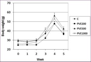 | Fig. 1- Body weight of mice receiving palm vitamin E supplementation before pregnancy Data is presented as mean ± SEM. C- control PVE200- palm vitamin E 200 mg/kg PVE500- palm vitamin E 500 mg/kg PVE1000- palm vitamin E 1000 mg/kg Week 0-2: mating and supplementation Week 2-5: gestational period and delivery |
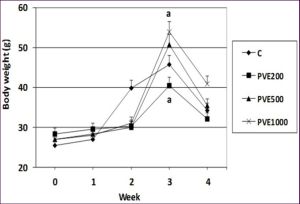 | Fig. 2- Body weight of mice receiving palm vitamin E supplementation during pregnancy Data is presented as mean ± SEM. Groups which share the same alphabet indicate significant difference (p<0.05). C - control PVE200- palm vitamin E 200 mg/kg PVE500- palm vitamin E 500 mg/kg PVE1000- palm vitamin E 1000 mg/kg Week 0-1: mating Week 1-4: gestational period, supplementation and delivery |
 | Table 1- Number, body weight and body length of pups from dams supplemented with palm vitamin E before pregnancy |
 | Table 2- Number, body weight and body length of pups from dams supplemented with palm vitamin E during pregnancy |