


ISSN : 2321-4430
EISSN :



SABNIS A.S.1*, JAMKAR A.A.2
1Department of Anatomy, K.J. Somaiya Medical College, Sion East, Sion, Mumbai- 400 022, MS, India.
2Department of Anatomy, S.B.H. Govt Medical College, Dhule- 424 001, MS, India.
* Corresponding Author : dranjus2003@yahoo.com
Received : 17-12-2012 Accepted : 01-01-2013 Published : 01-04-2013
Volume : 1 Issue : 1 Pages : 1 - 3
World Res J Anat 1.1 (2013):1-3
Conflict of Interest : None declared
<b>Background-</b> It is well known that palmaris longus (PL) is degenerating and variable muscle of flexor compartment of forearm. Distally at wrist joint it continues with palmar apponeurosis (PA) and strengthens it. Big tendon and small belly of PL fulfills the criteria for tendon transfer and donation. Variations in PL and in the formation of PA are clinically significant for hand surgeons. The study was done to find out the variations in PL & formation of PA and find out their incidence. <b>Methodology-</b> 60 upper limbs of 30 embalmed cadavers of both sex of age ranging between 60 to 80 years from the department of Anatomy were dissected to study the variations in PL and formation of PA. Measurement of total length of PL, tendon part and muscular part of PL muscle was measured. <b>Results</b> 1. In 11.6% of cases PL was absent unilaterally; 2. PA was present in all the cases; 3. In one palm PA is formed by tendon of Flexor Carpi Ulnaris in the absence of PL muscle; 4. In one palm PA is formed by duplicated PL along with separated thin tendon of Flexor Carpi Radialis. Conclusion- PA is always present. Presence of PA does not depend upon presence or absence of PL muscle. Though PL muscle is functionally less important it is useful for surgeries in hand. Variations in PL muscle carry clinical significance and such variations should not be ignored.
Palmaris Longus, Palmar Apponeurosis, Flexor carpi ulnaris
PL muscle is one of the superficial muscles in the flexor compartment of forearm lying between flexor carpi radialis laterally and flexor carpi ulnaris medially. It springs from medical epicondyle by the common tendon, adjacent intermuscular septa and deep fascia [1] . The tendon crosses flexor retinaculum and expands to form PA but few fibers of PL tendon incorporated into flexor retinaculum. PL muscle contracts palmar fascia and assists in flexing the wrist [2] . Absence of PL does not affect the functioning of wrist as other flexor muscles of forearm carry out flexion at wrist joint. Presence or absence of PL muscle does not affect the formation of PA. In absence of PL muscle PA is formed by other tendons like flexor carpi radialis or flexor carpi ulnaris and serves the function.
Such muscle having less function, small belly and long tendon is useful for orthopaedic surgeons in tendon transfer and graft.
The present study aimed to find out the incidence of any variation or absence of PL and formation of PA.
Dissection of 60 upper limbs of 30 embalmed cadavers of both sex, age ranging between 60 to 80 years from the department of Anatomy during the span of three years was carried out. Flexor compartment of forearm of both sides was dissected and all the flexor muscles were identified along with PL & PA. Measurement of total length of PL, tendon part and muscular part of PL muscle was measured by using divider and measuring scale.
PA formation was observed and any variation in it was noted.
Results
1. In 11.6% of cases PL was absent unilaterally.
2. PA was present in all the cases.
3. In one palm PA is formed by expansion of tendon of Flexor Carpi Ulnaris in the absence of PL muscle.
4. In one palm PA formation is formed by duplicated PL along with separated tendon of Flexor Carpi Radialis partly.
PL is considered as degenerating muscle as it has long tendon and short muscular belly. Distally it forms PA in palm which stabilizes the palmar skin which is firmly adhering to it [3] .
PA is triangular in shape and occupies central area of palm. The apex of PA is attached to distal border of flexor retinaculum and receives the insertion of PL tendon. The base of the apponeurosis divides at the bases of fingers into four slips. Each slip divides into two bands, one passing superficial to skin and other passing deep to root of finger and fuses with fibrous flexor sheath and deep transverse ligament. The function of PA is to give firm attachment to overlying skin and so improve the grip and to protect the underlying tendons [4] . Thus PL is helpful as flexor of wrist and stabilizer of palmar skin through PA. Absence or any variation of PL does not alter the function as the alternatives for carrying out flexion of wrist are available in the form of other flexors of forearm. Although PL muscle may be absent but PA is always present [5] . Various studies showed variable data related to unilateral or bilateral absence of PL [Table-1] .
We have found that PL was absent in 11.6% of cases unilaterally but PA was present in all the upper limbs. PA was attached to the flexor retinaculum proximally [Fig-1] and maintained normal anatomical structure distally. In none of the upper limbs bilateral absence of PL was seen.
In first case [Fig-2] where PL was absent, PA was formed by fibers of flexor carpi ulnaris, few fibers from flexor carpi ulnaris tendon traveled towards thinner eminence as well. On other side normal anatomy regarding PL & PA was present.
In second [Fig-3] case PL was duplicated having length of tendons 6 cm and 3 cm while length of muscle belly was 24 cm and 25 cm respectively. Both the tendons distally form PA in palm. One small and thin detached slip from flexor carpi radialis also took part in the formation of PA. Such rare formation of PA with duplication of PL is not documented yet. Duplication of PL was observed in one upper limb [13] .
In third case [Fig-4] palmaris longus tendon was bifurcated at two places & forms PA. The one slip given proximally reunites with the main tendon and distal slip goes to thinar eminence and got incorporated with fascia covering it. Flexor carpi ulnaris was inserted on pisiform bone, but just before insertion small part of tendon traveled towards flexor retinaculum & takes part in its formation.
We have measured the length of PL muscle, tendinous and muscular part of PL muscle. [Table-2] .
PL is commonly used by hand surgeons for tendon transfer [12] . It is first choice donor tendon as it fulfils the necessary requirements of length, diameter and availability and can be used without producing any functional deformity [16] . Any accessory PL muscle that appeared to compress the ulnar nerve during repeated contractions [17] .
Precise anatomy of PA is important for understanding the basis of Dupuytren’s contraction in which progressive shortening of PA causes flexion of fingers. Surgical division of PA is required for straightening it. Usually PA is formed by PL tendon expansion in palm. Variations in the formation of PA by flexor carpi ulnaris, flexor carpi radialis, and duplication is not yet documented. Awareness of such variations should not be ignored during surgical correction in hand surgeries.
1. PA is always present though variations are seen in structures forming PA.
2. Greater importance should be given to PA, PL tendon and their variations by hand surgeons.
[1] Standring S. (2008) Gray’s Anatomy, 40th ed., London, Churchill Livingstone Elsevier.
» CrossRef » Google Scholar » PubMed » DOAJ » CAS » Scopus
[2] Salter M.I. (1987) Hand Injuries-A Therapeutic Approach, 1st ed., Edinburg, London, Churchill Livingstone.
» CrossRef » Google Scholar » PubMed » DOAJ » CAS » Scopus
[3] Romanes G.J. (2003) Cunningham’s Manual of Practical Anatomy, 1, 15th ed., United States, Oxford University.
» CrossRef » Google Scholar » PubMed » DOAJ » CAS » Scopus
[4] Snell R.S. (2000) Clinical Anatomy for Medical Students, 6th ed., Philadelphia, Lippincott Williams & Wilkins.
» CrossRef » Google Scholar » PubMed » DOAJ » CAS » Scopus
[5] Tank P.W. (2008) Grant’s Dissector, 14th ed., Philadelphia, Lippincott Williams & Wilkins.
» CrossRef » Google Scholar » PubMed » DOAJ » CAS » Scopus
[6] Stecco C. (2009) Clinical Anat., 22(2), 221-9.
» CrossRef » Google Scholar » PubMed » DOAJ » CAS » Scopus
[7] Caughell K.A. (1988) J. Hand Surg. Am.,13(4), 485-93.
» CrossRef » Google Scholar » PubMed » DOAJ » CAS » Scopus
[8] Agarwal P. (2010) Indian Journal of Orthopaedics, (44)2, 212-215.
» CrossRef » Google Scholar » PubMed » DOAJ » CAS » Scopus
[9] Kapoor S.K. (2008) Anat. Sci. Int., 83(1), 45-48.
» CrossRef » Google Scholar » PubMed » DOAJ » CAS » Scopus
[10] Eric M. (2010) Surg. Radiol. Anat., 32(4), 357-361.
» CrossRef » Google Scholar » PubMed » DOAJ » CAS » Scopus
[11] Sater M.S. (2010) Clin. Anat., 232(8), 956-961.
» CrossRef » Google Scholar » PubMed » DOAJ » CAS » Scopus
[12] Roohi S.A. (2007) Malaysian Orthopaedic Journal, 1(1), 26-28.
» CrossRef » Google Scholar » PubMed » DOAJ » CAS » Scopus
[13] Mangala M. (2008) Romanian Journal of Morphology and Embryology, 49(2), 215-217.
» CrossRef » Google Scholar » PubMed » DOAJ » CAS » Scopus
[14] Mbaka G.O. (2009) Ulster Med. J., 78(2), 90-93.
» CrossRef » Google Scholar » PubMed » DOAJ » CAS » Scopus
[15] Thompson N.W. (2001) The Ulster Medical Journal, 70(1), 22-24.
» CrossRef » Google Scholar » PubMed » DOAJ » CAS » Scopus
[16] Troha F. (1990) Ann. Plast. Surg., 25(6),477-478.
» CrossRef » Google Scholar » PubMed » DOAJ » CAS » Scopus
[17] Robert P.H. (1972) Hand, 4(1), 40-41.
» CrossRef » Google Scholar » PubMed » DOAJ » CAS » Scopus
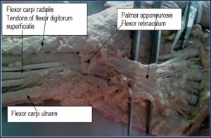 | Fig. 1- PA attached to the Flexor Retinaculum proximally |
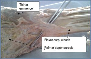 | Fig. 2- Absence of PL, PA was formed by fibers of flexor carpi ulnaris, few fibers from flexor carpi ulnaris tendon traveled towards thinner eminence |
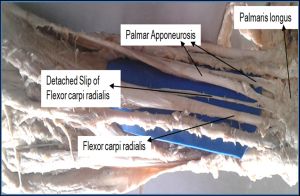 | Fig. 3- PL was bifurcated distally, PA was formed by bifurcated PL tendon & thin detached slip from flexor carpi radialis |
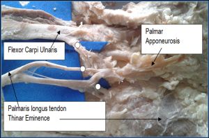 | Fig. 4- Distal bifurcation of flexor carpi ulnaris & PL tendon |
 | <i>Table 1- Shows results of the study done by other authors</i> |
 | <i>Table 2- Shows measurement of PL in present study compared with study done by Stecco</i> |