


ISSN : 0976-7061
EISSN : 0976-707X



SABNIS A.S.1*
1Department of Anatomy, K.J. Somaiya Medical College, Sion, Mumbai-400022, MS, India
* Corresponding Author : dranjus2003@yahoo.com
Received : 29-11-2012 Accepted : 04-12-2012 Published : 24-12-2012
Volume : 3 Issue : 2 Pages : 46 - 48
J Clin Res Lett 3.2 (2012):46-48
DOI : http://dx.doi.org/10.9735/0976-7061.3.2.46-48
Conflict of Interest : None declared
Background: Sciatic nerve (SN) arises from sacral plexus, leaves the pelvis and enters in gluteal region via greater sciatic foramen. It divides into tibial (TN) and common peroneal nerve (CPN), usually in popliteal fossa. This point of bifurcation may be variable and it is clinically important. Compression of the nerve at any point during its course may give rise to pain. Difference in clinical symptoms is observed depending upon the compression point of the nerve. So the study aimed to investigate the variations in bifurcation point of sciatic nerve. Methodology: 140 lower extremities of 70 cadavers of age 50-70 years of both sex during 5 years were studied to see the variations in sciatic nerve bifurcation.All cadavers were embalmed with 10% of formalin. The gluteal region was dissected well and sciatic nerve course was studied by noting the point of bifurcation. Results: In the first type higher bifurcation of sciatic nerve below piriformis found in 7% of cases while in second type we found bilateral higher bifurcation was in one cadaver. Conclusion: Though sciatic bifurcation at lower level either in popliteal fossa or upper 1/3 of thigh is common, awareness of higher bifurcation and their relation to piriformis, obturator internus, gamelli muscle is clinically important.
Sciatic Nerve, Tibial Nerve, Common Peroneal Nerve, Piriformis Muscle, Obturator Internus Muscle.
Sciatic, the thickest nerve in the human body is a branch of sacral plexus (L4, L5, S1, S2 & S3). From pelvis via greater sciatic foramen it enters in gluteal region by passing inferior to piriformis. It descends between greater trochanter and ischial tuberocity along the back of thigh. It has two components, TN ((L4, L5, S1, S2 & S3) & CPN (L4, L5, S1, S2) into which it bifurcates in the popliteal fossa usually [1] .
Variations are seen in the point of bifurcation unilaterally or bilaterally. In the higher bifurcation one of the nerve i.e TN or CPN may get spared in case of injury to sciatic nerve. Though complete palsy of sciatic nerve is rare to occur it results into flail foot and severe difficulty in walking [1] . Anatomical variations may contribute to piriformis syndrome, sciatica, coccygodynia and muscle atrophy [2] . Awareness of higher or lower, unilateral and bilateral variations in point of bifurcation carries clinical significance. The study was conducted to study the variations in the point of bifurcation and to find out the incidence of different types of bifurcation.
70 embalmed cadavers of both sex between the ages of 30-70 years were obtained from department of anatomy of K.J. Somaiya & Seth G.S. Medical college in the span of 7 years. Gluteal region of both the lower extremities was dissected by reflecting skin, fat, and gluteus maximus muscle. Sciatic nerve, piriformis, obturator internus along with two gamelli and quadratus femoris was identified. The course of sciatic nerve was studied in detail & the point of bifurcation was noted.
• Majority (90.7%) of lower limbs (LL) show bifurcation of sciatic nerve at lower level [Table-1] and 9.28% of LL show at higher level [Table-2] .
• In only one cadaver bilateral higher bifurcation was seen, one side shows division below piriformis and other side shows piriformis in between CPN & TN.
• CPN was entering in gluteal region by piercing through obturator internus muscle in one case & in between obturator internus and piriformis muscle in another case.
The origin of word sciatic is of Latin origin from Greek word “ischiadikos†which means subject to trouble in the hips or loins [3] . It is the thickest nerve of the body and nerve of posterior compartment of the thigh. It is formed in the pelvis from lumbar and sacral nerves and enters in gluteal region through greater sciatic foramen below piriformis. The nerve passes along the back of thigh where it is crossed by biceps femoris and divides into tibial and common peroneal nerve in the popliteal fossa [1] . Variations are seen regarding the bifurcation of sciatic nerve at different levels. This bifurcation can be at lower level i.e. in popliteal fossa or posterior compartment of thigh or at upper level i.e. in gluteal region.
Various studies [Table-3] have reported different levels of bifurcation of sciatic nerve. In our study we have found bifurcation in popliteal fossa in 24% of lower extremities unilaterally [Fig-1] & [Table-1] , 34% bilaterally & in upper 1/3rd of thigh in 16% unilaterally [Fig-2] & [Table-1] , 15% bilaterally. Incidence of bifurcation of sciatic nerve in popliteal fossa is 36% [4] , 67% [5] , 32% [6] , at apex of popliteal fossa in 46.67%, below apex in 33.3% [7] . Sciatic nerve divides at lower 2/3rd of femur in 13.8% of cases [4] ., in middle third of thigh in 10.4% of cases [5] , above apex of popliteal fossa in 20% of cases [7] .
Regarding upper level division we have observed four different types of divisions in relation to piriformis, obturator internus & gamelli muscle.
In the first type [Fig-3] & [Table-2] we have found higher bifurcation of sciatic nerve below piriformis in 7% of cases. Such higher level bifurcation can result into involvement of only one out of two divisions resulting in a decrease in neurological deficit as compared to low bifurcation of sciatic nerve & failure of popliteal block anaesthesia [8] . We did not find any case showing CPN piercing piriformis muscle.
In second type, [Fig-4] & [Table-2] bilateral higher bifurcation was seen in one cadaver. On left side sciatic nerve division was seen below piriformis and on other side CPN was seen superior to piriformis & TN was seen inferior to piriformis. Piriformis is present in between the two nerves. Such incidence is 8% [9] . In 10% of cases the two divisions exit the pelvis as distinct nerves. In these situations TN always passes below the piriformis muscle while CPN can pass above or through piriformis. The CPN is more prone to mechanical injury than the TN because it has larger and more tightly packed funiculi with less protective connective tissue and because of its more lateral position and tethering in sciatic notch and head of fibula [13] .
In [Fig-4] on right side two branches of sciatic nerves are separated by piriformis muscle & on left side bifurcation occurs below piriformis muscle.
In third case [Fig-5] & [Table-2] CPN entered in gluteal region by piercing obturator internus muscle & TN was seen inferior to Inferior Gamellus muscle. Such piercing of obturator internus is not yet documented.
In fourth case [Fig-6] & [Table-2] CPN was seen passing between piriformis & obturator internus with two gamelli & TN was seen inferior to inferior Gamellus muscle. Such variation is very rare. Mas et al have also reported a case of bilateral high division of sciatic nerve but TN was passing under superior gamellus [14] .
In the whole course of sciatic nerve from sacral plexus to popliteal fossa, bifurcation of sciatic nerve can occur at any point which carries clinical significance. Bilateral neuropathy or trauma of sciatic nerve is rare to occur but unilateral lesion of sciatic nerve is common due to compression, trauma & injury during hip replacement procedure. Any variation regarding point of bifurcation, course of sciatic, CPN, TN may aggravate the compression of nerve and gives rise to pain in lower limb. To find out exact cause of pain, knowledge regarding anatomical variations in sciatic nerve, CPN, TN will be helpful in treating the patients.
Such high bifurcation motivates radiologist to repeat MRI on other side, as there can be difference on two sides [12] . Awareness of presence of such variations will be significant for surgeons, intern doctors, general practioners, nurses to achieve required goals.
[1] Susan Standring (2008) Grays Anatomy, 40th ed., London, Churchill Livingstone Elsevier.
» CrossRef » Google Scholar » PubMed » DOAJ » CAS » Scopus
[2] Babinski M.A., Machado F.A., Costa W.S. (2003) Eur. J. Morphol., 41, 41-2.
» CrossRef » Google Scholar » PubMed » DOAJ » CAS » Scopus
[3] Sarita S. (2012) Anat. Physiol., 2(5), 1-4.
» CrossRef » Google Scholar » PubMed » DOAJ » CAS » Scopus
[4] Okraszewska E. (2002) Folia Morphol, 61, 4, 277-282.
» CrossRef » Google Scholar » PubMed » DOAJ » CAS » Scopus
[5] Ogeng’O J.A. (2011) Folia Morphol, 70, (3), 175-179.
» CrossRef » Google Scholar » PubMed » DOAJ » CAS » Scopus
[6] Muthu Kumar T., (2011) J. of Clinical and Diagnostic Research, 5(8), 1502-1504.
» CrossRef » Google Scholar » PubMed » DOAJ » CAS » Scopus
[7] Sherry Sharma (2012) J.K. Practitioner, 17, 1-3.
» CrossRef » Google Scholar » PubMed » DOAJ » CAS » Scopus
[8] Prakash (2010) Singapore Med. J., 51(9), 721-723.
» CrossRef » Google Scholar » PubMed » DOAJ » CAS » Scopus
[9] Siyatik S. (2009) Turkish Neurosurgery, 19(2), 139-144.
» CrossRef » Google Scholar » PubMed » DOAJ » CAS » Scopus
[10] Chiba S. (1992) Kaibogaku Zasshi, 67(6), 691-724.
» CrossRef » Google Scholar » PubMed » DOAJ » CAS » Scopus
[11] Machado F.A. (2003) Int. J. Morphol., 21(1), 29-35.
» CrossRef » Google Scholar » PubMed » DOAJ » CAS » Scopus
[12] Patel S. (2011) National J. of Medical Research, 1(2).
» CrossRef » Google Scholar » PubMed » DOAJ » CAS » Scopus
[13] Stuart L. Weinstein, Joseph A. Buckwalter (2005) Turek’s Orthopaedics Principles & their Application, 6th ed., 555.
» CrossRef » Google Scholar » PubMed » DOAJ » CAS » Scopus
[14] Mas N., Ozdemir B. (2003) Neuroanatomy, 2, 13-15.
» CrossRef » Google Scholar » PubMed » DOAJ » CAS » Scopus
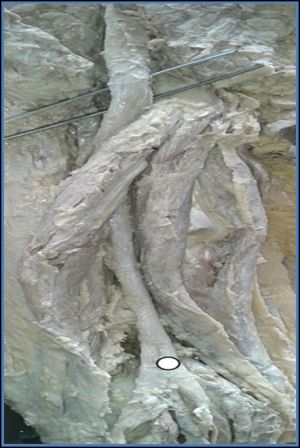 | Fig. 1- Sciatic bifurcation in popliteal fossa |
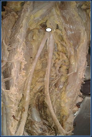 | Fig.2- Sciatic bifurcation at upper 1/3rd of thigh |
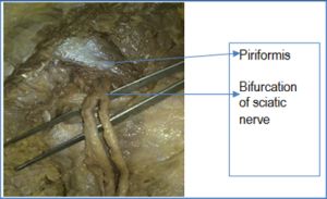 | Fig. 3- Unilateral higher bifurcation of sciatic nerve below piriformis |
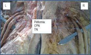 | Fig. 4- Bilateral higher bifurcation of sciatic nerve. |
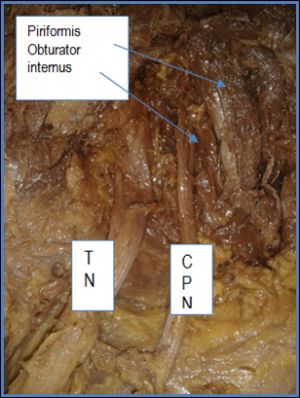 | Fig. 5- Higher bifurcation of sciatic nerve, CPN coming out through obturator internus muscle. |
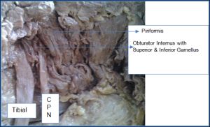 | Fig. 6- CPN passing between piriformis and obturator internus with two gamelli & TN passing inferior to inferior gamellus |
 | Table 1- Bifurcation of sciatic nerve at lower level |
 | Table 2- Bifurcation of sciatic nerve at higher level |
 | Table 3- Results of study on sciatic bifurcation at higher level done by several authors |