


ISSN : 0976-8726
EISSN : 0976-8734



FLANNERY M.T.1*, LINETTE RIVERA2, BEATA CASANAS3
1University of South Florida College of Medicine, 17 Davis Blvd., Room 208, Tampa, FL 33606
2University of South Florida College of Medicine, 17 Davis Blvd., Room 208, Tampa, FL 33606
3University of South Florida College of Medicine, 17 Davis Blvd., Room 208, Tampa, FL 33606
* Corresponding Author : mflanner@health.usf.edu
Received : 22-03-2012 Accepted : 12-04-2012 Published : 03-12-2012
Volume : 3 Issue : 1 Pages : 23 - 25
Med Case Rep 3.1 (2012):23-25
DOI : http://dx.doi.org/10.9735/0976-8726.3.1.23-25
Conflict of Interest : None declared
Cryptococcal infections in human hosts typically present with pulmonary, cutaneous or central nervous system involvement. The patient presented with upper thoracic back pain with a osteolytic lesion of the second thoracic vertebrae suggestive of neoplasm. A biopsy and serology confirmed the infection was cryptococcal neoformans. Our patient developed acute tubular necrosis and a type I distal RTA secondary to intravenous liposomal amphotericin requiring a change to high dose intravenous fluconazole followed by a six months course of oral fluconazole therapy.
Cryptococcus, Osteomylitis, Acute Tubular Necrosis, Distal RTA, Fluconazole.
A sixty five year old Latino female presented with a one month history of worsening right scapular and shoulder pain that increased with movement and had awakened her from sleep. She denied a history of fever or chills, night sweats, weight loss, a rash or any neurologic complaints. The patient had an eight year history of type II diabetes mellitus that had been well controlled with metformin 500mg three times daily. She had recently been using hydrocodone with acetaminophen as needed for the right scapular and shoulder pain.
A mammogram two months prior to admission was normal; but she had no other preventive health care performed. Her surgical history consisted of only a cholecystectomy at the age of thirty as well as a bilateral tubal ligation at age twenty nine. She was a native of Honduras but had lived in the United States for many years. She had recently visited Honduras for two weeks approximately three months prior to admission; she denied any illness while visiting Honduras.
There was no alcohol or tobacco use or known risk factors for the Human Immunodeficiency Virus (HIV). Household animals included a family parrot and several pigeons that she had owned for many years.
A physical examination revealed that she was afebrile with normal vital signs; her Body Mass Index (BMI) was thirty two. She had small freely moveable anterior cervical adenopathy on the right with a right thyroid nodule of approximately two centimeters. The remainder of the examination was unremarkable.
Laboratory data indicated a white blood cell count of 7.6 mm [3] , hemoglobin 12.7 g/dl with a platelet count of 297/mm3; the differential was unremarkable. Electrolytes and renal function were normal and the initial fasting glucose was 225 mg/dl. The hemoglobin A1c was 8.7%. The remainder of her metabolic profile was normal except for an albumin of 3.5 g/dl. A chest radiograph demonstrated a questionable increase in the right paratracheal density. Computed tomography (CT) of the chest and abdomen demonstrated a 2 x 1.3 cm subpleural nodule at the right lung base with central cavitation. It also demonstrated multiple lymph nodes in the aorta/pulmonary window, left hilar and right paratracheal area that were subcentimeter in size. The lung parenchyma revealed scattered peripheral irregular septal thickening with areas of nodularity and confluent ground-glass opacities within the right upper lobe, lingula of the left lobe and bilateral lower lobes. There was a lytic lesion involving the T2 vertebral body extending into the ventral epidural space without cord compression. The imaging of abdomen and pelvis revealed no masses or adenopathy.
Magnetic resonance imaging of the neck and cervical/thoracic spine demonstrated an abnormality in the T2 vertebral body, predominantly on the right side and involving the right pedicle without evidence of cord compression. There was some abnormal soft tissue signal in the right paravertebral region, as well as, an enlarged thyroid gland with heterogeneity. Additional laboratory studies showed an erythrocyte sedimentation rate of 34 mm/hr. A serum protein electrophoresis demonstrated a mild polyclonal gammopathy. Elisa testing for human immunodeficiency virus 1 and 2 were negative. The absolute CD 4 count was 852 mm [3] with a normal CD4 to CD8 T-cell ratio. A thyroid stimulating hormone level was normal. A lumbar puncture was performed and the cerebral spinal fluid (CSF) revealed no nucleated cells with a normal glucose, VDRL negative and a CSF total protein of 47mg/dl. Gram stain, acid fast bacillus and fungal smears and cultures were all negative. The India ink and CSF cryptococcal antigen were also negative.
A CT guided biopsy of the T2 vertebral body was obtained which demonstrated PAS fungal and GMS fungal stains consistent with cryptococcus neoformans and no evidence of malignancy. A serum cryptococcal antigen was positive at 1:155. A positron emission tomography scan showed intense activity in T2, with non-specific activity in the lungs and bowel loops. A thyroid scan and uptake could not be performed due to the recent administration of intravenous contrast for computed tomography.
The patient was immediately administered liposomal amphotericin B as well as flucytosine, but after one week developed distal renal tubular acidosis and worsening renal insufficiency. She was subsequently switched to intravenous high dose fluconazole. The right scapular pain gradually improved with this agent as well as analgesics and physical therapy. She was subsequently discharged to finish a two week course of intravenous fluconazole followed by oral fluconazole for a total of six months.
Cryptococcus neoformans was first described in 1984 as an encapsulated yeast which reproduces by budding and forms round yeast-like cells that are 3 to 6 um in diameter. There are two varieties of Cryptococcus: neoformans and gattii. The species has four serotypes based on the antigenetic specificity of the capsular polysaccharide. Cryptococcus neoformans var. neoformans is often recovered from pigeon feces, bird nests, guano and is invariably serotypes A or D. Cryptococcus neoformans can be cultured with the addition of creatinine as a nitrogen source, which partly explains the growth of this organism in creatinine-rich avian feces. Cryptococcus neoformans var. neoformans causes the vast majority of cryptococcal human infections in immunosuppressed hosts including patients with HIV/AIDS whereas cryptococcus neoformans var. gattii causes seventy to eighty percent of all cryptococcal infections among immunocompetent hosts [1] .
Following inhalation, the yeast deposits in the pulmonary alveola; the host response to subsequent cryptococcal infection includes both cellular and humoral components. Bone involvement develops in five to ten percent of patients and is usually osteolytic or resembles cold abscesses. These lesions may be confused with tuberculosis or neoplasm [2] . A review of the general features of forty cases of cryptococcal osteomyelitis demonstrated that the majority of infections occurred in patients twenty nine to fifty nine years of age with a slight male predominance. Single site involvement occurred in seventy five percent of the patients, whereas twenty five percent had multiple sites of involvement [3] .
Risk of infection is most commonly seen in the vertebrae (twenty five percent) and less commonly in the femur, tibia and rib (17.5 percent [3] . Recent case reports also demonstrate involvement of the calvarium, digits and clavicle [4-11] . Underlying diseases most commonly included sarcoidosis in twenty five percent followed by tuberculosis in 12.5 percent, steroid therapy in ten percent and diabetes melitis in 7.5 percent of all cases [3] . The pathogenesis of cryptococcal osteomyelitis is typically hematogenous dissemination from a pulmonary focus or lymph node or direct inoculation of the skin with contiguous spread. In those patients who have evidence of cord compression surgical decompression is peremptory [3] .
In spite of the cautious implementation of a liposomal amphotericin B preparation, the patient nevertheless developed acute tubular necrosis and type I distal renal tubular acidosis. Subsequently, the patient received a course of high dose intravenous fluconazole for two weeks, to be followed with a course of additional oral therapy for a total of 6 months.
The updated guidelines from the Infectious Disease Society of America in 2010 recommended that non-meningeal non-pulmonary cryptococcosis with infection limited to a single site may be treated with fluconazole [6mg/kg per day] for 6-12 months [12] . Several case reports published over the last decade have demonstrated successful treatment of cryptococcal osteomyelitis with fluconazole alone [8,10-17] . Many of these cases had successful treatment outcomes with three months of fluconazole therapy; however due to the significant pulmonary involvement presented by our patient, a more prolonged course of fluconazole was warranted.
In general, patients with evidence of cryptococcal infections should undergo a lumbar puncture to ascertain the absence of CNS infection. Clinical and radiographic follow-up as well as cryptococcal serum titers should be monitored to ensure a favorable response to anti-fungal therapy [6] .
Our case was typical in that the patient was older with underlying diabetes and presented with a pulmonary focus and a history of bird exposure. The biopsy was diagnostic as was the markedly elevated serum cryptococcal antigen. Lack of renal tolerance to amphotericin B warranted an alternative treatment, so the patient was subsequently switched to fluconazole therapy. Although it is rare, cryptococcal osteomyelitis should be considered in patients with an underlying chronic disease, exposure to bird excrement and lytic bone lesions. It is anticipated that she will recover completely from her disease after six months of fluconazole therapy.
[1] King J.W., Cunha B.A. (2011) eMedicine Inf Dis.
» CrossRef » Google Scholar » PubMed » DOAJ » CAS » Scopus
[2] Goldshteyn N.A., Zanchi A., Cooke K., Agha R. (2006) SMJ, 9, 1140-1141.
» CrossRef » Google Scholar » PubMed » DOAJ » CAS » Scopus
[3] Yuk-Fong Liu P. (1998) Diagn. Microbiol. Infect. Dis., 30(31), 33-35.
» CrossRef » Google Scholar » PubMed » DOAJ » CAS » Scopus
[4] Ching N., Lasky J., Lazareff J., Moore T.B., Nielsen K. (2004) Ped. Inf. Dis. J., 3(6), 589.
» CrossRef » Google Scholar » PubMed » DOAJ » CAS » Scopus
[5] Rocco A., Fleckenstein J., Brandvold B., Ondra S. (1993) Neurosurgery, 32(6), 1034-36.
» CrossRef » Google Scholar » PubMed » DOAJ » CAS » Scopus
[6] Hawkings C., Flaherty J. (2007) Inf. Dis. in Clin. Prac., 15(5), 345-47.
» CrossRef » Google Scholar » PubMed » DOAJ » CAS » Scopus
[7] Corral J., Lima S., Quezada J., Samayoa B., Arathoon E. (2011) Med. Mycol., 49(6), 667-71.
» CrossRef » Google Scholar » PubMed » DOAJ » CAS » Scopus
[8] Mruthyunjaya J., Ravishankar R. (2011) Indian J. Pathol. Microbiol., 54(1), 216-18.
» CrossRef » Google Scholar » PubMed » DOAJ » CAS » Scopus
[9] Agadi J.B., Madni N.A., Nanjappa V., Govindaiah H.K. (2010) Neurology India., 58(2), 300-2.
» CrossRef » Google Scholar » PubMed » DOAJ » CAS » Scopus
[10] Amit A., Sudish K., Pople I.K. (2008) Acta Neurochirurgiaca, 150(7), 713-14.
» CrossRef » Google Scholar » PubMed » DOAJ » CAS » Scopus
[11] Karmakar P., Jeyarajih R., Ramasubramanian V. (2011) J. Indian Med. Assoc., 109(8), 592-94.
» CrossRef » Google Scholar » PubMed » DOAJ » CAS » Scopus
[12] Perfect J.R., Dismukes W.E., Dromer F., Goldman D.L., Graybill J.R., Hamill R.J., Harrison T.S., Larsen R.A., Lotholary O., Nguyen M.H., Pappas P.G., Powderly, W.G., Singh, N., Sobel J.D., Sorrell T.C. (2010) Clinical Infect Dis., 50, 291-322.
» CrossRef » Google Scholar » PubMed » DOAJ » CAS » Scopus
[13] Cook P. (2001) SMJ, 94(9), 936-38.
» CrossRef » Google Scholar » PubMed » DOAJ » CAS » Scopus
[14] Chang W.C., Tzao C.H., Hsian-He C., Chang H., Lo C.P., Chen C.Y. (2005) J. Infect., 51(3), e117-19.
» CrossRef » Google Scholar » PubMed » DOAJ » CAS » Scopus
[15] Murphy S., Parnell N. (2005) J. Infect., 51(5), e309-11.
» CrossRef » Google Scholar » PubMed » DOAJ » CAS » Scopus
[16] Al-Tawfiq J., Ghandour J. (2007) Inf, 35(5), 377-82.
» CrossRef » Google Scholar » PubMed » DOAJ » CAS » Scopus
[17] Irfan Q., Farheen A., Umair Z., Masood U. (2011) J. Infect Dev Ctries, 5(9), 669-73.
» CrossRef » Google Scholar » PubMed » DOAJ » CAS » Scopus
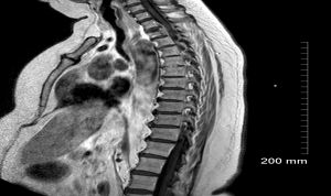 | Fig. 1- MRI spine demonstrating lytic lesion of T2 vertebral body. |